SiO2/(��-Fe2O3-SiO2)���Դ���������Ʊ�
������, ��ʢ��, ������, �쾰��
(�廪��ѧ ����������Դ�����о�Ժ, ���� 100084)
ժ Ҫ: �����ܽ�-�������Ʊ��˾��и����ȶ��ԵĦ�-Fe2O3-SiO2���ϲ���, ������������SiO2����, ��������׳ߴ�ź˵ĵ���ɢ����SiO2/(��-Fe2O3-SiO2)������ X������������� ���ز��ȷ����� ɨ��羵�� ��羵�Լ�����Ʒ��ǿ�Ƶ�ʵ����˵��: Fe2O3-SiO2�����ᆳ��700�������¶�����30min��ų��֦�-Fe2O3����, �����¶Ȼ��߸���������ʱ�佫���¦�-Fe2O3���-Fe2O3��ת��; ��SiO2������õ���SiO2/(��-Fe2O3-SiO2)Ϊ�������ȵĵ���ɢ���ο���, �����ߴ���150~200nm����, �ڲ�����Ϊ��-Fe2O3����, ��������ܵ�SiO2������ ͨ������Fe3O4�����ֱ�Ӱ���SiO2���Ա�ʵ�����, SiO2/(��-Fe2O3-SiO2)���и�������Ĵ�����, ��һ�������Ĵ����������塣
�ؼ���: ��-Fe2O3; ���Դ�������; ���ȶ���; ���� ��ͼ�����: TQ13
���ױ�ʶ��: A
Preparation of SiO2/(��-Fe2O3-SiO2) magnetic catalyst carrier
WANG Song-wei, XU Sheng-ming, CHEN Song-zhe, XU Jing-ming
(Institute of Nuclear and New Energy Technology,Tsinghua University, Beijing 100084, China)
Abstract: Fe2O3-SiO2 composite was prepared with sol-gel method. SiO2/(��-Fe2O3-SiO2) magnetic catalyst carriers were obtained by coating composite with silica. These obtained samples were characterized by XRD, TG-DTA, TEM, SEM and VSM. The effects of processing temperature, time and initial dosage of iron nitrate on the properties of Fe2O3-SiO2 were studied. The results show that, after calcined at 700�� for 30min, the composite oxide containing ��-Fe2O3 phase is secured. While further increase of the processing temperature (>800��) or time results in the formation of ��-Fe2O3 phase. SEM and TEM analyses show that the SiO2/(��-Fe2O3-SiO2) sample is of nano-sized, monodisperse spherical particles with size of 150-200nm, which are well coated by amorphous SiO2 layer. The VSM data exhibit that SiO2/(��-Fe2O3-SiO2) has much better magnetic properties than SiO2/Fe3O4. Such nano-composites are very promising in application as magnetic catalyst carriers and sorbent carriers.
Key words: ��-Fe2O3; magnetic catalyst carrier; heat-resistance; coating
��ϸ�Լ����׳ߴ�Ĵ������ھ߱��ȱ������ ����λ�ḻ���ص�, ������������ڴ�ͳ�Ĵ�ߴ����, ���ֳ����Ӧ�ü�ֵ�� �������������Ӧ���д������ѷ��������, ������������˷Ѻ����ʹ�óɱ�, ͬʱ��������Ϊ�ŷŶ�������Ⱦ�� ���ʹ���������Ͼ߱�����, �������ʹ�ú�ͨ����Ӵų�ʵ�ּ���Ч�ķ��롣 Ŀǰ�����Ѿ���ʼ���Զ��ִ�������Ϊ����, ���������������ʵġ����Դ��������о�[1, 2], ����Ϊһ����д���Ӧ���ԵĴ���, ����˴��Բ��Ϻʹ���������, �ڹ�������� �������� ��ת�ƴ��� ����� �������� ������������ھ������õ�Ӧ��ǰ��[3, 4]�� ��������������������������(Fe3O4)�� ��-����������(��-Fe2O3)����ɱ�����, �����ܽϺ�, ����ѡ�����Դ������ں�, �÷��澡�����н϶���о���չ[5-8], ��Ŀǰ��˵, ���ǻ������Դ�������������(Fe3O4)���-����������(��-Fe2O3)����Ϊ����, �����Ʊ���ϸ����������ʱ����������һЩ����, ���͵���: 1)��ϸ������Fe3O4���ߦ�-Fe2O3�����к�ǿ���ž�����, ���ѿ��ƶ���ֱ�Ӱ����õ����ղ���Ŀ�����״�� ������С���ֲ�[9-11]; 2)�ں�̵����������ʰ���������, ������Ҫ��������(�����ܽ�-�������������ѿ���TiO2ʱ, ��Ҫ400�����ϵ������¶�), ��ʱ�����ź˵����ȶ������Բ���, ��: Fe3O4���ߦ�-Fe2O3��������������������, ��400�������¶��¼���ת��Ϊ�����ԵĦ�-Fe2O3[12], ���´��Դ��, ʹ����ʧȥ�ɴ��Ի��յ����ԡ�
�������߲����ܽ�-�������Ʊ��˦�-Fe2O3-SiO2������, SiO2�ĸ�������˦�-Fe2O3�����ȶ���, ��Ч�����������-Fe2O3��ת��[13-15], Ȼ���Դ˸�����Ϊ�����ں�, �����ϰ���SiO2��, �����ö�ΰ����ķ�������������������ֲ�, ���ջ�������������Ҿ��нϸ����ȶ��Ե���״���Դ�������SiO2/(��-Fe2O3-SiO2)��
1 ʵ��
1.1 ��-Fe2O3-SiO2��������Ʊ�
������������(������)�� ��ˮ�Ҵ�(������)�� ȥ����ˮ��Ħ����Ϊ1��3.85��10.2���Ƴ���Һ, ����Fe2O3��(Fe2O3+SiO2)��Ħ���ȷ�Χ0.071~0.350, ��һ������Fe(NO3)3��9H2O������Һ��, ����1h, ��������ҺpHֵ��0.7�����Եõ����ȶ����ܽ�, �ڿ�������Ȼ������, �����������ں����л���������110��õ��������� ��ĥ��ֱ��ڲ�ͬ�¶�������(300~900��)���յõ�Fe2O3-SiO2�����
1.2 SiO2/(��-Fe2O3-SiO2)���Ʊ�
��1.0g �������������������40mL��ˮ�Ҵ��в���������30min, ����20mL������������Һ(4mL����������+16mL��ˮ�Ҵ�), ����μ���һ������ˮ��Һ, ���������·�Ӧ6h�Ա�֤����������ˮ����ȫ, ���Ĺ��˺�Ӧ�������110�������º��, �õ�SiO2/(��-Fe2O3-SiO2)���Դ������塣 �ظ��˲��輴�ɽ��ж��ΰ���, �Ե���SiO2������ĺ���Լ������Ĵ�С��
Ϊ�����Ա�ʵ��, ������Fe3O4(ALDRICH��ѧҩƷ��˾��, ����С��5��m, ����98%)��Ϊ�����ں�, ������ͬ��ʵ��������輰ҩƷ��������SiO2����, ���յõ�SiO2/Fe3O4��
1.3 ��Ʒ�ı���
��Ʒ��X��������������ձ���ѧ D/max-RB��X�����������Ͻ���, �������Ϊ: Cu K��, ��ѹ40kV, ����100mA; ��NETZSCH STA 409C�Ͳ����������Ϸ�����(��������20��/min)��Fe2O3-SiO2����������չ��̽�������-���ȷ���ʵ��(TG-DTA); Fe2O3-SiO2�������SiO2/(��-Fe2O3-SiO2)���Դ����������ò������Amary 1910FE��ɨ��羵(SEM)���м��, �ڲ��ṹ����JEM-2010�߷ֱ����������(HRTEM, ���ٵ�ѹ200.0kV)��JEM-1200EX�����������(TEM, ���ٵ�ѹ120.0kV)���м�⡣ ��������LakeShore 7307������Ʒ��ǿ��(VSM)����Ʒ���д��Բ�����
2 ���������
2.1 ��-Fe2O3-SiO2����������ʱ���
��-Fe2O3-SiO2�����������Ʊ����̵�����-���ȷ��������ͼ1��ʾ�� TG���߱��������������������330��ʱ������ʧ�Ѿ�������ȫ; DTA��������ʾ, ��Ʒ��650�����ҿ�ʼ, �����˻��������ȹ���, ��Ϧ�-Fe2O3-SiO2�������X����������(ͼ2)��֪, �����ȹ��̶�Ӧ�ڸ���������������Fe2O3���γɦ�-Fe2O3����, �������-Fe2O3����ת��Ĺ��̡�
��-Fe2O3-SiO2��������Fe2O3��Ħ������Ϊ0.188, ����ʱ��Ϊ0.5h��������, ������ͬ�������¶�(500~ 900��)���պ�����Fe2O3-SiO2��X������������ͼ2��ʾ�� ��ͼ��֪, ��500��600��������ʱ, ������û���γ�Fe2O3����, ֱ��700��ʱ�ų��֦�-Fe2O3����������, 800������ͬʱ���֦�-Fe2O3�ͦ�-Fe2O3����������ź�, ����500 ~ 900������������¾���������������(���������)�� �ɴ˿�֪, SiO2�ĸ����ܹ�������߸�����������Fe2O3������¶�, ��ܿ����Ǹ�
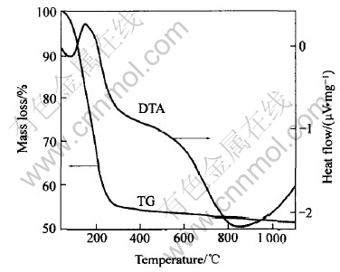
ͼ1 ��-Fe2O3-SiO2�����������-��������
Fig.1 TG-DTA curves of precursor of ��-Fe2O3-SiO2 sample
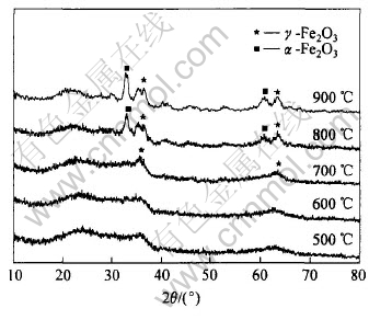
ͼ2 ����ͬ�¶����պ��-Fe2O3-SiO2�������X����������
Fig.2 XRD patterns of ��-Fe2O3-SiO2 samples heat treated at different temperatures
��������״�ṹ��SiO2�ӻ���Fe2O3�ľ����Լ���һ���ľ���ת��, ���ۻ��������һ��̽�֡�
Ϊ��������ʱ���Ӱ��, ��x=0.188, �����¶�700���������, �Ը������ֱ������0.5, 1.0��2.0h������, ��X������������ͼ3��ʾ�� ����0.5h�����ո�������ֻ�����˦�-Fe2O3����, ����������ʱ����ӳ�, �����֦�-Fe2O3���岢�����ࡣ ͼ4��ʾΪt=0.5h, ��=700��ʱ, �ڲ�ͬ��xֵ(����ͬFe2O3����)������������Ʒ��X���������ס� �ɿ�����������Fe2O3��������ʱ�ܹ������֦�-Fe2O3����, ��Fe2O3�������ӵ�һ���̶�ʱ(x=0.279), ������Ʒ�л�ͬʱ���֦úͦ�-Fe2O3, Ҳ����˵, ���Ÿ�������Fe2O3����������, ��-Fe2O3���-Fe2O3���ת���¶Ƚ��ή�͡�
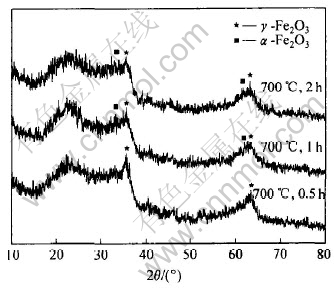
ͼ3 ��ͬ����������Fe2O3-SiO2��Ʒ��X����������
Fig.3 XRD patterns of Fe2O3-SiO2 samples heat treated under different conditions
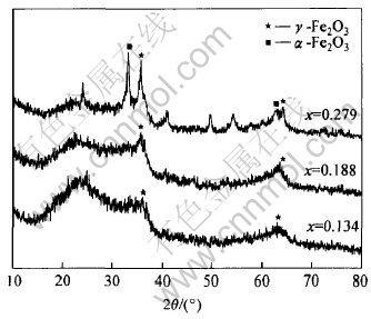
ͼ4 ��ͬFe2O3����ʱFe2O3-SiO2��Ʒ�����պ��X����������
Fig.4 XRD patterns of Fe2O3-SiO2 samples with different contents of Fe2O3 after heat-treatment
2.2 SiO2/(��-Fe2O3-SiO2)������
ͼ5��ʾΪx=0.188, 700������0.5h�����������õĦ�-Fe2O3-SiO2���������SiO2ǰ���ɨ��羵��Ƭ�� ���Կ���, ����ǰ��������״�ܲ�����, �����ֲ�Ҳ�ܲ�����, ����������SiO2������, ���õ���SiO2/(��-Fe2O3-SiO2)�����ѱ����״��Ϊ����ĵ���ɢ���ο���, �����ߴ���150~200nm���ҡ� ��-Fe2O3-SiO2��SiO2/(��-Fe2O3-SiO2)�������ڲ��ṹ��羵��Ƭ��ͼ6��ʾ�� ͼ6(a)��ʾΪx=0.188, 700������0.5 hʱ���õĦ�-Fe2O3-SiO2���������羵��Ƭ, ���Ц�-Fe2O3(ͼ�гĶȽ������)��SiO2���ҵ��ս���һ��, ���֦�-Fe2O3������¶���ⲿ; ����������SiO2������õ���SiO2/(��-Fe2O3-SiO2)����, ��ͼ6(b)��ʾ, �������ȫ�����ܵ�SiO2��������, �ڲ�������һ���ϴ�Ħ�-Fe2O3-SiO2������������С�Ħ�-Fe2O3-SiO2������ ͼ6(c)��ʾΪ�Ա�ʵ���д�Fe3O4��������SiO2���������õ���SiO2/ Fe3O4����, ���Կ�������Fe3O4�ں˿�����С��һ, ����SiO2/ Fe3O4������״�ϲ�����, �����ֲ�������, ͬʱSiO2�������Ȳ�����, �Ҵ��ھֲ�û�а�����ȫ������ ����SEM��TEM�������, ��ʵ����Ʊ����������ܹ��Ϻõ����SiO2�Ԧ�-Fe2O3-SiO2������İ���, �������Ҫ�Ĺ���ĵ���ɢ���ο���; ����һ����, SiO2/(��-Fe2O3-SiO2)������, �����Ӧ�-Fe2O3�����в�����, ����չ����һ����ʵ�鹤������������ơ�
2.3 �����ܱ���
�Ԧ�-Fe2O3-SiO2(x=0.188, 700������0.5h)������SiO2�����õ�SiO2/(��-Fe2O3-SiO2) ��������Ʒ��ǿ����(VSM), �����ͼ7��ʾ�� ͼ�л������˶�����ƷSiO2/ Fe3O4(700������0.5h)��VSM���Խ���� ���Կ���, �����Ż���ǿ��, SiO2/ Fe3O4�Ѵﵽ����, ����-Fe2O3-SiO2��SiO2/(��-Fe2O3-SiO2)��δ�ﵽ����, ������8.0��105A/m�ĴŻ���ǿ��, ��-Fe2O3-SiO2�Ĵž�(1.58A��m2)��SiO2/(��-Fe2O3-SiO2)�Ĵž�(0.89A��m2)����SiO2/ Fe3O4�Ĵž�(0.08A��m2)Ҫ��ö�, ������Ϊ��700�����պ�, SiO2/(��-Fe2O3-SiO2)�еĦ�-Fe2O3����δ�����ı�, ��SiO2/ Fe3O4��Fe3O4ת��Ϊ��-Fe2O3����Ӷ���
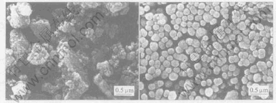
ͼ5 ��-Fe2O3-SiO2����SiO2ǰ(a)�Ͱ�����(b)��ɨ��羵��Ƭ
Fig.5 SEM images of ��-Fe2O3-SiO2 before(a) and after(b) coating
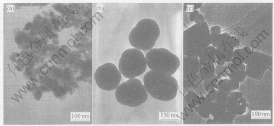
ͼ6 ��-Fe2O3-SiO2����SiO2ǰ(a)�� ������(b)��Fe3O4����SiO2��(c)����羵��Ƭ
Fig.6 TEM images of ��-Fe2O3-SiO2 before coating (a),��-Fe2O3-SiO2 after coating (b) and Fe3O4 after coating (c)
�´��Դ��[12]�� ������ڱ�ʵ��������, �������������õ�SiO2/(��-Fe2O3-SiO2)����Ĵ���Ҫ����ֱ�ӽ�SiO2�����ڴ�Fe3O4�����ò���Ĵ��ԡ�
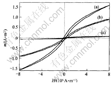
ͼ7 ��-Fe2O3-SiO2����SiO2ǰ(a)�� ������(b)��Fe3O4����SiO2��(c)������Ʒ���ͻ���
Fig.7 VSM data of ��-Fe2O3-SiO2 before coating (a), ��-Fe2O3-SiO2 after coating (b) and Fe3O4 after coating (c)
3 ����
ͨ����Fe2O3Ԥ����SiO2�Ʊ��ɸ���������, �ܹ���Ч������Fe2O3�����, ��ø����ȶ��ԵĦ�-Fe2O3-SiO2, �ڴ˻����Ͻ���SiO2����, �ɹ��ػ���˵���ɢ����SiO2/(��-Fe2O3-SiO2)������ ���ڸÿ����ܹ��ڽϸ��¶��±�������Fe2O3���Ϊ����, ά�������, ������ڲ������շ������Ͻ��н�һ���Ļ�����ָ���, �Ӷ��õ����Դ���, ��˱�ʵ�����Ƴ���SiO2/(��-Fe2O3-SiO2)������һ�������Ĵ����������塣
REFERENCES
[1]Watson S, Beydoun D, Amal R. Synthesis of a novel magnetic photocatalyst by direct deposition of nanosized TiO2 crystals onto a magnetic core[J]. Journal of Photochemistry and Photobiology A: Chemistry, 2002, 148: 303-313.
[2]Beydoun D, Amal R, Low G, et al. Occurrence and prevention of photodissolution at the phase junction of magnetite and titanium dioxide[J]. J Mole Catal A: Chem, 2002, 180: 193-200.
[3]Nixon L, Koval C A, Noble R D, et al. Preparation and characterization of novel magnetite-coated ion-exchange particles[J]. Chem Mater, 1992, 4: 117-121.
[4]Pierre A, Vladimir S, Richard J, et al. Preparation and properties of magnetite and polymer magnetite nanoparticles[J]. Langmuir, 1999, 15: 1945-1951.
[5]Redl F X, Cho K S, Murray C B, et al. Three-dimensional binary superlattices of magnetic nanocrystals and semiconductor quantum dots[J]. Letters to Nature, 2003, 423: 968-971.
[6]Larken E, Stephaine G, O��Brien S, et al. Cooperative assembly of magnetic nanoparticles and block copolypeptides in aqueous media[J]. Nano Letters, 2003, 3: 1489-1493.
[7]David I, Frank C. Spontaneous phase transfer of nanoparticulate metals from organic to aqueous media[J]. Angew Chem Int Ed, 2001, 40(16): 3001-3004.
[8]Patricia B, Nicholas B, Katie J, et al. Preparation and properties of an aqueous ferrofluid[J]. Journal of Chemical Education,1999, 76(7): 943-948.
[9]Liu Q X, Xu Zh H, Finch J A, et al. A novel two-step silica-coating process for engineering magnetic nanocomposites[J].Chem Mater, 1998, 10: 3936-3940.
[10]Albert P, Michel P B, Chellapah P. Magnetic silica dispersions: preparation and stability of surface-modified silica particles with a magnetic core[J]. Langmuir, 1994, 10: 92-99.
[11]Taeghwan H, Su S L, Jongnam P, et al. Synthesis of highly crystalline and monodisperse maghemite nanocrystallites without a size-selection process[J]. J Am Chem Soc, 2001, 123: 12798-12801.
[12]������, ���Ӿ�, ������, ��. �Ʊ��������������������͵�Ӱ��[J]. ��ըҩѧ��, 2002, 4: 85-86.
LIU Xiang-xuan, WANG Xuan-jun, LIU Zhen-yu, et al. Effect of synthesis method on crystal phase of iron oxide nanocrystalline powders[J]. Chinese Journal of Explosives & Propellants, 2002, 4: 85-86.
[13]Ennas G, Musinu A, Piccaluga G, et al. Characterization of iron oxide nanoparticles in an Fe2O3-SiO2 composite prepared by a sol-gel method[J]. Chem Mater, 1998, 10: 495-502.
[14]Cannas C, Concas G, Falqui A, et al. Investigation of the precursors of ��-Fe2O3 in Fe2O3/SiO2 nanocomposites obtained through sol-gel[J]. Journal of Non-Crystalline Solids, 2001, 286: 64-73.
[15]Cannas C, Musinu G, Piccaluga G. Structural investigation of Fe2O3-SiO2 nanocomposites through radial distribution functions analysis[J]. Phys Chem Chem Phys, 2004, 6: 3530-3534.
(�༭������)
������Ŀ: ������Ȼ��ѧ����������Ŀ(50274045)
�ո�����: 2005-07-15; ������: 2005-08-20
�����: ������(1980-), ��, ��ʿ�о���
ͨѶ����: ��ʢ��, ������; �绰: 010-89796082; ����: 010-62773585; E-mail: smxu@ tsinghua.edu.cn

