���±�ţ�1004-0609(2011)12-3175-07
������ʴ�����и�����ͭ����ò�仯��Ԫ��Ǩ��
�� ��1��������1����ݼع1, 2
(1. ����������ѧ ���Ͽ�ѧ�빤��ѧԺ������ 100029��
2. �й��Ļ��Ų��о�Ժ ������ѧ�����о��������� 100029)
ժ Ҫ�������ں�ģ����������(0.010 4 mol/L Na2SO4+0.028 2 mol/L NaCl+0.016 4 mol/L NaHCO3)�����������������ͭ�����ķ����о�������ͭ��������ʴ���ɣ����ý��������۲츯ʴǰ����ͭ�Ľṹ�仯��ɨ��羵(SEM)���������(EDS)�����ɷֽ��з�������X����ӫ�����(XRF)������������Χ������Ԫ�غ�����ͬʱ�ò�������fCu/Sn��fCu/Pb����������ʴ������Cu��Sn��Pb�IJ������������������ڸû����¸�����ͭ��ʴ���ȴӦ����������濪ʼ���Ҧ������ڦ������ʴ������δ��ʴ�ĵ���״���ࣻ������ͭ��ʴ������Cu��������Χ������Ǩ�ƣ������γɵĺ�O��C��ʴ�����и���Sn��Pb��
�ؼ��ʣ�������ͭ����������ʴ��Ǩ��
��ͼ����ţ�TG 174 �� �� ���ױ�־�룺A
Morphology change and elements migration of bronze with high tin content after soil corrosion
TANG Qi1, WANG Ju-lin1, MA Jing-yu1, 2
(1. School of Materials Science and Engineering, Beijing University of Chemical Technology, Beijing 100029, China;
2. Protection Institute of Science and Technology, Chinese Academy of Cultural Heritage, Beijing 100029, China)
Abstract: The soil corrosion rule of the bronze with high tin content was researched by burying the metal samples in the soil containing the simulated the edaphic elecrdyte (0.010 4 mol/L Na2SO4+0.028 2 mol/L NaCl+0.016 4 mol/L NaHCO3) aimed at simulating the underground water. The morphology change before and after the corrosion was observed by metalloscopy, and SEM combined with EDS was used to analyze the micro-area composition of the corroded sample. Further research with X-ray fluorescence spectrometry (XRF) was carried out to measure the content of the elements in the soil around the bronze sample. Then, factors fCu/Sn and fCu/Pb were used to make a quantitative analysis of the residual elements Cu, Sn and Pb in the corrosion products. The results show that the corrosion firstly occurs on the interface between the �� phase and �� phase, �� phase is prior to be corroded, with the island-shaped �� phase uncorroded and left. Cu is prior to diffuse into the surrounding soil, leaving the corrosion products containing O and C rich in Sn and Pb.
Key words: High-tin bronze; soil; corrosion; diffusion
����ͭ�Ͻ�ʴ���ɵ��о����������ﱣ��ѧҲ�ǽ�����ʴѧ�о������һ�����ſ��⡣���͵�������ͭ��֯�ɦ��������(��+��)���������[1-4]���й���ͭ��֯�ṹ�Ц���ͦ�������ȸ�ʴ���������Ѿã��е���Ϊ�Ǧ���(��ͭ��)���ȸ�ʴ[5-6]���е���Ϊ���༰����(������)����һ�����ȸ�ʴȡ�������ĺ���[7]�������о�����[3, 8-9]����ͭ���港ʴ�����и������ɴ��ƶ���ͭ����(������)������ͭ��ʴ�γɸ�ʴ�������ͭ����������ʴ����ͭ��ʧ�������������ʵ�ϣ����۲쵽�ĸ�ʴ���������ܽ�Ľ������Ӵӻ�������Ǩ�ơ���������ʧ����������Ԫ������Ǩ�ƣ������������γɵ�[10]�����ԣ�����ͨ��������ͭ���港ʴ�������ƶϸ�ʴ������ԭ�����Է�ӳ��ʴ��������ʵ���̡�
�Խ�ɳ��ַ������ͭ������ɷֺͼ�̬���еķ����������Գ��������������������SnO2��ʹ����ͭ���������һ����ʴ[11]��������4������ս��ʱ����ͭ����ì�ļ���з��ִ���SnO2Ϳ�㣬������������ͭì����Ϳ��һ������������Ϳ���Է���ʴ������֤������������Ϳ��ķ���Ч�����[12]��
������������������о��丯ʴ���̺�����һ����Ҫ��������ʴ�о�������ʵ����ɿ����ܹ���һ���̶���ģ��������ʴ����ʵ�����������ӵ�����ϵͳʹ�ø������غܶ࣬�����˶Խ���������Ѷȡ�Ȼ������ģ�����������н���ʵ����ʧȥ������������������ʵ�ԡ�Ϊ�˾����ܽ��������������ĸ����ԺͲ��ɿ����ԣ���ʵ�齫�ɷֺ�Ũ����֪��ģ����ʼ��뵽��ϴ���������У�ͨ���ں�ģ���������ʵ������������ͭ�Լ���ģ���������ͭ������ʴ���̣�����ͭ��ʴǰ�������֯���ɷֱ仯�����������治ͬ���봦����������Ԫ�����༰�京���仯���ϣ��Ӹ����Ͻ�ʾ��ͭ��ʴ�����Լ�������Ҫ����Ԫ�ص�Ǩ�ƹ��ɡ�
1 ʵ��
ʵ���ø�����ͭ�����Dz��ô�ͳ���취����ұ���������Cu-Sn-Pb��Ԫ�Ͻ𣬴�ĥ������XRF��������(����������%)���£�Cu 72.8%��Sn 23.0%��Pb 4.2%���������и��10 mm��10 mm��3 mm�ķ��飬���ͣ��û�����֬��̣�������200#��800#ˮɰֽ��ĥ�����棬��ȥ����ˮ��ϴ���ƾ����ú��ﱸ�á�
ʵ��������ȡ�Ա��������Ÿ���һʩ��������Լ10 m����������Ա���������и�ֳ���������ʴ���ܵ�Ӱ�졣�����ʵ��������������ʡ��ƺ�ɫ��ͬʱ��Ϊ�˾����ܽ���������ԭ�����ڵĿ��������ʶ�ʵ��ĸ��ţ�������������ˮ��ϴ3�飬ȥ����ˮ��ϴ3�飬���ɷ�����ÿ�ԼΪ1 mm�IJ���������ˣ�ȡ�²�ϸ����Ϊʵ�������õ��������������о�������pHֵΪ8.05�ĺ�0.010 4 mol/L Na2SO4+ 0.028 2 mol/L NaCl+0.016 4 mol/L NaHCO3��ģ�����ˮ��Һ[13-14]���ο�ʪ�ȶ�X70�ָ�ʴ����Ӱ��Ĺ���[15]��������Ƴɺ�ˮ��30%�ĸ�ʴ��������
������ճ�������Ƴ��ڳ����β�����(25 cm��5 cm��5 cm)��һ�ˣ���ÿ�����м������úõĸ�ʴ������900 g������ñ���Ĥ��ס�����ۿڡ�ÿ��7 d�ҿ�����Ĥһ�Σ����ڷ���Լ30 min���ÿ������롣�����ȿ��Ա�������ʪ���ֿ���ʹ�������룬ģ�⺬�������³�ʪ��������ͭ�ĸ�ʴ���ɡ������120 d������ȡ����ͬʱ�ֱ��ھ�����������0��2��4��6�� 8 cm��ȡ�ߴ�ԼΪ1 cm��1 cm��0.5 cm�������飬�������������ԡ�
1) ��������֯�۲�
��ëˢ��ȥ��ʴ����ͭ���������������ʴ���Ϊ�˲��ƻ���ʴ�����֯��̬������������������¹۲졣ͬʱ����δ�����������ʵ�����ͭ��������֯���й۲���Ϊ�Աȣ������ǰ���ͭ����������200~2000��ˮɰֽ��ĥ������2.5 ��m���ʯ��ĥ���⣬ȥ����ˮ��ϴ����3%FeCl3�ƾ���Һ��ʴ������ȥ����ˮ��ϴ��������������¹۲졣����Ϊ�¹�Leica��˾������Leica DM4000 M��ѧ������
2) SEM-EDS���ɷֲ���
�������Ƕ�ɨ��羵�¹۲쵽�ĸ���ͬ����ɷֽ��в��ԡ������ͺż������������£�������˾S-3600N��ɨ��羵�����ٵ�ѹ20 kV����Ʒ�õ��罺ճ����Ʒ̨�Ϲ۲졣����EDAX��˾DX-100��X��������ɫɢ���ǣ���������Ϊ��գ�������ѹΪ15 kV��
3) XRF����
���ձ�����˾������EDX-800HS X����ӫ������Ƕ���ȡ�����ɷֽ��в��ԡ���������Ϊ��գ�����ԴΪ���(Rh)��������ѹ��Ti-U 50 kV��Na-Sc 15 kV��
2 ���������
2.1 ��֯�ṹ���ɷֱ仯
SCOTT[16]������������ͬ���Ŵ�����ͭ��Ϊ�����͵������࣬������С��17%��Ϊ������ͭ����ʵ����������ͭ������Ϊ23.0%�����ڸ�����ͭ��δ��غ���������Ľ�����֯��ͼ1��2��ʾ����ͼ1�ɿ��������������ͭ���и�����ͭ���͵Ħ��������(��+��)��������֯�ṹ������Cu-Sn��Ԫ�Ͻ���ͼ(��ͼ3)[17]��ͼ1�к����ϸߵ�Ϊ(��+��)�����塣ͬʱ���������ֲ�������̬��Ǧ�������һЩ�������ɡ�����
�ں���ģ���������ʵ����������120 d����ͭ�����淢�������ظ�ʴ����֯��ò�����˱仯(��ͼ2)���ڸ�ʴ��̫���ص�����(��ͼ2(a)��(b))�����Գ����ó���ʴ�����������ų�������֯��(��+��)������߽���У����ҷ����ڳ�������֯�ڡ��ڸ�ʴ�Ƚ����ص�����(��ͼ2(c)��(d))�����Գ����ó��ʵ���״�ĺ�ɫ��ʴ�����DZ�(��+��)����������Χ���ѻ���������ȫ��ʴ�ij������࣬����֦״�ĺ�ɫ��ʴ������(��+��)�������й����Ħ��ฯʴ״����(��+��)�������ڵĦ������û�з�����ʴ��
Ϊ�˽�һ��ȷ�����ȸ�ʴ���Ǧ���Ǧ��࣬��SEM���ҵ�������ͼ2(d)��ʾ������(��ͼ4(a))������ͼ�а�������������������ɫ����ɫ����ɫ����ֱ��������ǽ��гɷַ����������ͼ4(b)��(c)��(d)��ʾ����3��������Cu��Sn��Pb 3��Ԫ�ص�����������Ħ���������бȽϣ�������1���С�
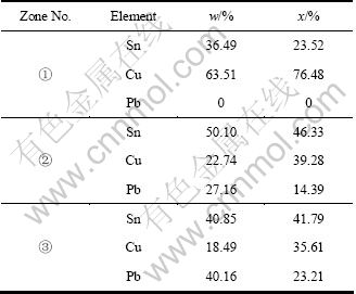
����١���SEM�������ڷ���ǿ����������ɫ���京������ò������(��+��)��������δ������ʴ�Ħ��࣬EDS���Խ������������ٳ�������C��N��OԪ���⣬��Ҫ��Cu��Sn����Ԫ����ɡ���1�м�������������������Cu��SnĦ����Ϊ31:9.5������ͼ3�ĺϽ���ͼ���������Խ����仯����Cu31Sn8Ϊ����Ĺ����壬��Cu��Sn������Ħ����ԼΪ31:8������������ijɷּ������dz��ӽ������Զ϶�������Ϊ�������еĦ�����������δ������ʴ��

ͼ1 FeCl3��Һ��ʴ�����ͭ����Ľ�����֯
Fig.1 Metallographs of bronze base after corrosion by FeCl3 solution
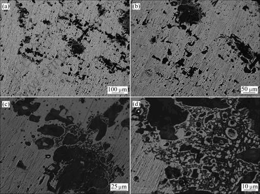
ͼ2 ������ʴ����ͭ�����Ľ�����֯
Fig.2 Microstructures of bronze sample after buried in soil
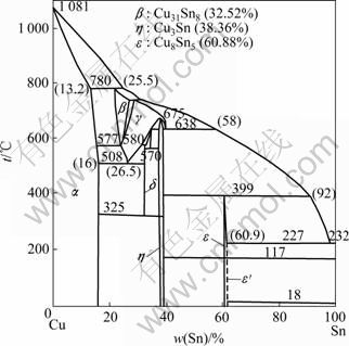
ͼ3 Cu-Sn��Ԫ�Ͻ���ͼ
Fig.3 Phase diagram of Cu-Sn alloy
����ڡ���SEM����Ϊ��ɫ������Ƭ����EDS���Խ����ʾ��Cu��SnĦ����Ϊ0.85������ͼ3��������Sn�ĺ���Ϊ15.8%(��������)[17]����Cu��SnĦ����ԼΪ10����ˣ����������Ǧ���ijɷֱȽϣ��������ò������ڸ�ʴ��ʹCuǨ����ʧ���أ�����Cu������ԭ���������Խ��ͣ���Sn��Pb�ĺ������Ƚϸߣ�������Sn��O�ĺ���Ҳ�ܸߣ������γ��˺����ĸ�ʴ�������丯ʴ��ò������ȷ��������Ǧ�����˸�ʴ��
����ۡ�EDS���Խ��������ڵ����ơ�Cu��Sn��Ħ����ԼΪ0.85��Ҳ�Ǧ������ʴ������������ɫ����ò������������������𣬶��Ҹò�����Pb��C����Ҫ������ڵĸߣ����ԣ�����Pb��C��ʴ���������ڵĸߡ�
���������������������120 d�ĸ�����ͭ����ʴ���Ŧ���ͦ���Ľ�����У��Ҧ���Ȧ������ȷ�����ʴ����������[18-20]�н��ܵļ����������ͭ�Ͻ����ʴ�ԡ����߸�����ͭ�ȵ�����ͭ��ʴ��һ�µġ�
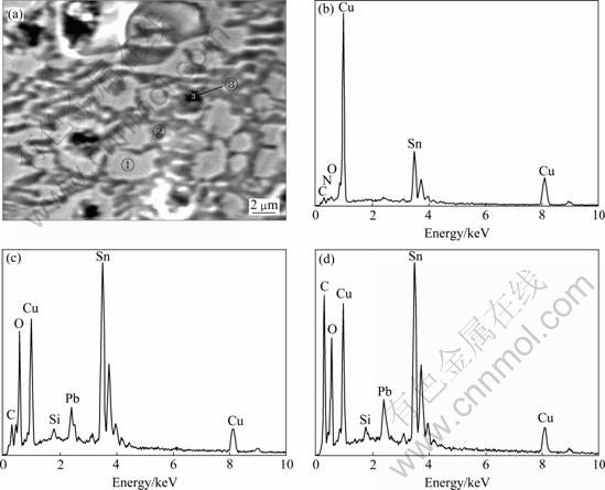
ͼ4 ������ͭ��ʴ�����SEM������EDS�������
Fig.4 SEM image of bronze sample after buried and EDS analysis results: (a) SEM image; (b) Zone ��; (c) Zone ��; (d) Zone ��
��1 �������������
Table 1 EDS analysis results of micro-zones
2.2 ��ͭ�Ͻ��Ԫ�ص��ܽ⡢Ǩ�Ƽ�����
ͨ�������������治ͬλ�ƴ���������Ԫ�ؼ��京���仯�����Ի����ͭ�Ͻ�Ԫ�ص�Ǩ�ƹ��ɡ���2�����������������120 d�ĸ�����ͭ������ͬ���봦Ԫ�غ�������仯��������6 cm��8 cm��û�м�Cu��Sn��Pb���κ�һ��Ԫ�أ�����ֻ�г���0 cm��2 cm��4 cm�ļ����������CuԪ���Ѿ�����Χ������Ǩ��������4 cm������0~8 cm��Χ�ڲ�û�з���Sn��PbԪ�ء��������EDS���������ʴ������Sn��Pb�����ܸߣ���CuԪ�غ����Ȧ���ͦ����еĵͺܶ࣬˵��Cu�����Sn��Pb���ڴӸ�ʴ��������(�˴�Ϊ����)����Ǩ�ơ�
��2 �����ͭ��Χ�����ɷּ����������
Table 2 Composition and content of soil around buried bronze

Ϊ�˸�����ؽ�ʾ������ͭ��������ʴ����и�Ԫ�������ܽ⡢Ǩ�ƺ������ʴ�����״���������ܽ����ӵĸ���[9, 19, 21-22]���ܽ�����fCu/Sn��fCu/Pb��ʾ��ʴ������Cu��Sn��PbԪ�ص��ܽ⡢Ǩ�ƺ������ʴ����������
��ͼ4(a)�и�ʴ����ڡ��������������Ϊԭʼ���ݽ��м��㡣
 (1)
(1)
 (2)
(2)
ʽ�У�wCu,p��wSn,p��wPb,p�ֱ��ʾ��ʴ������Cu��Sn��PbԪ�ص�����������wCu,a��wSn,a��wPb,a�ֱ��ʾ��ͭ�Ͻ�(��+��)��������֯������Cu��Sn��PbԪ�ص������������۲����������ͭ������֯���ֺ�Ǧ���������ϼ�����(��+��)�������У�ͬʱ���躬Ǧ����ȫ���ֲ��ڹ�����Ħ����У�����������Cu��Sn��PbԪ�ص����������ֱ�Ϊ72.63%��13.64%��13.73%��
��fCu/Sn��fCu/Pb=1ʱ���þ��봦�����ĸ�ʴ������w(Cu):w(Sn)��w(Cu):w(Pb)��Ͻ��е���ͬ���� 0��fCu/Sn��fCu/Pb��1ʱ���þ��봦�����ĸ�ʴ������w(Cu):w(Sn)��w(Cu):w(Pb)С�ںϽ��еģ�˵���ô�Sn(��Pb)��ʴ��������ı����ϴ�Cu�����Sn(��Pb)ѡ���Ը�ʴ������Ǩ�Ƴ�ȥ����fCu/Sn��fCu/Pb��1ʱ���þ��봦������ʴ������w(Cu):w(Sn)��w(Cu):w(Pb)���ںϽ��еģ�˵���ô���ʴ����Cu�����ı����ϴ�Sn(��Pb)�����Cuѡ���Ը�ʴ������Ǩ�Ƴ�ȥ�����ݼ��������3��ʾ��
�ӱ�3���Կ�������ͬ��ʴ����IJ�������fCu/Sn��fCu/Pb��С��1���Ҳ��ּ������ӽ���0��˵����ʴ������Sn��Pb�����ı����ܴ�ʴ���︻Sn��Pb��������Cu�����Sn��Pb����Ǩ�Ƴ�ȥ�������������еIJ������������ġ�
��3 ��ʴ����IJ�������fCu/Sn��fCu/Pb
Table 3 Residual factor of fCu/Sn and fCu/Pb in corrosion products

3 ����
1) ������ͭ�ں���0.010 4 mol/L Na2SO4+0.028 2 mol/L NaCl+0.016 4 mol/L NaHCO3ģ�����ˮ��Һ�����������120 d��ʴ���Ŧ���ͦ���Ľ�����У����Ҧ���Ȧ������ȷ�����ʴ����ʴ������Sn��Pb��Cu�ĺ�O��C���������δ������ʴ��
2) �����120 d�������ͭ����0~4 cm�������У�ֻ������Cu��Sn��Pb 3��Ԫ�أ�����CuԪ���Ѿ�����Χ������Ǩ��������4 cm��˵��Cu�ĸ�ʴ���������Sn��Pb�ĸ�ʴ������״Ӹ�ʴ������Ǩ�ơ�
3) ��ʴ����IJ�������fCu/Sn��fCu/Pb��С��1���Ҳ��ּ������ӽ���0��˵����ʴ������Sn��Pb�����ı����ܴ�ʴ���︻Sn��Pb��������Cu�����Sn��Pb����Ǩ�Ƴ�ȥ��
REFERENCES
[1] ����÷, ԭ˼ѵ, �� ��, �ܱ���. ��ԭ��ַ�����Ĺ�س�����ͭ����ʴ�о�[J]. ���ﱣ���뿼�ſ�ѧ, 1999, 11(2): 7-18.
ZHANG Xiao-mei, YUAN Si-xun, LIU Yu, ZHOU Bao-zhong. Research on the corrosion of bronzes from Zhouyuan site and Yu state cemeteries[J]. Sciences of Conservation and Archaeology, 1999, 11(2): 7-18.
[2] ����ͭ�ӹ������������ҽ�����. ͭ��ͭ�Ͻ������ͼ[M]. ����: ұ��ҵ������, 1983.
Metallurgical Group of Luoyang Copper Processing Factory Central Laboratory. Diagrams of copper and its alloy[M]. Beijing: Metallurgical Industry Press, 1983.
[3] �� ��, ԭ˼ѵ, ����÷. ����-�����ܴ�����Ĺ�س�����ͭ����ʴ�о�[J]. ���ﱣ���뿼�ſ�ѧ, 2000, 12(2): 9-18.
LIU Yu, YUAN Si-xun, ZHANG Xiao-mei. Research on the corrosion of bronze wares excavated from Tianma-Qucun site of Jin state in Zhou Dynasty[J]. Sciences of Conservation and Archaeology, 2000, 12(2): 9-18.
[4] ��С��, ������, ���վ�. �ȴ�����Ǧ����ͭ��ʴ���ܵ�Ӱ��[J]. �й���ʴ�����ѧ��, 2008, 28(2): 112-115.
FAN Xiao-pan, WANG Chang-sui, JIN Pu-jun. Effect of heat treatment on corrosion behavior of bronze[J]. Journal of Chinese Society for Corrosion and Protection, 2008, 28(2): 112-115.
[5] �����, �� �, �Ʒﴺ, ������, ������. ����ʡ������������ͭ����ʴ�����о�[J]. ��ʴ��ѧ���������, 2007, 19(3): 157-161.
LUO Wu-gan, QIN Ying, HUANG Feng-chun, HU Ya-li, WANG Chang-sui. Study on corrosion products of some amcient bronzes excacated from Hubei province[J]. Corrosion Science and Protection Technology, 2007, 19(3): 157-161.
[6] �� ��, �λ���, ������, Ф �U. ģ����ͭ����Ʒ�ڵ��͵������Һ�еĵ绯ѧ��Ϊ�о�[J]. ���ﱣ���뿼�ſ�ѧ, 2007, 19(4): 45-48.
WANG Ning, HE Ji-quan, SUN Shu-yun, XIAO Lin. Bronze samples in various typical electrolytes[J]. Sciences of Conservation and Archaeology, 2007, 19(4): 45-48.
[7] SCOTT D A. Bronze disease: A reviews of some chemical problems and the role of relative humidity[J]. Journal of the American Institute for Conservation, 1990, 29: 193-206.
[8] SCOTT D A. An examination of the patina and corrosion morphology of some Roman bronzes[J]. J Am Institute Conserv, 1994, 33(1): 1-23.
[9] SIDOT E, SOUISSI N, BOUSSELMI L, TRIKI E, ROBBIOLA L. Study of the corrosion behaviour of Cu-10Sn bronze in aerated Na2SO4 aqueous solution[J]. Corrosion Science, 2006, 48: 2241-2257.
[10] ROBBIOLA L, TRAN T T M, DUBOT P, MAJERUS O, RAHMOUNI K. Characterisation of anodic layers on Cu-10Sn bronze (RDE) in aerated NaCl solution[J]. Corrosion Science, 2008, 50: 2205-2215.
[11] ���ƻ�, ��˼ά, �� ��. ��ͭ����Ĺ����������[J]. ���ϱ���, 2007, 40(2): 67-70.
CHEN Shan-hua, LIU Si-wei, SUN Jie. Photoelectron spectroscopy analysis of broze artifacts[J]. Materials Protection, 2007, 40(2): 67-70.
[12] �� Ⱥ, ������, ������, ���. ������ͭ��������������EPMA�о�[J]. ��ɢ��ѧ��, 2005, 17(2): 192-199.
YANG Qun, WANG Yi-lin, ZHANG Peng-xiang, LI Chao-zhen. The corrosion study of Raman spectra EPMA of bronze at Yunnan province[J]. Chinese Journal of Light Scattering, 2005, 17(2): 192-199.
[13] WANG Ju-lin, XU Chun-chun, L? Guo-cheng. Formation of CuCl and regenerated Cu crystals on bronze surfaces in neutral and acidic media[J]. Applied Surface Science, 2006, 252: 6294-6303.
[14] ASTMD 1384��01. Standard test method for corrosion test for engine coolants in glassware [S]. 2001.
[15] ��С��, ������, ����÷, ����ǿ, �����. ʪ�ȶ�X70������ʯ�������и�ʴ��ΪӰ��ĵ绯ѧ�о�[J]. ��ʴ��ѧ���������, 2007, 27(1): 35-37.
FEI Xiao-dan, LI Ming-qi, XU Hong-mei, LI Yong-qiang, CHAI Duo-chang. Influence of soil humidity on corrosion behavior of X70 steel in yellow pebble soil[J]. Corrosion Science and Protection Technology, 2007, 27(1): 35-37.
[16] SCOTT D A. Metallography and microstructure of ancient and historic metals[M]. London: The Getty Conservation Institute in Association with Archetype Books, 1991.
[17] �� ǿ. ������ɫ��������ͼ�״�ȫ[M]. ����: ұ��ҵ������, 2005: 886-889.
GAO Qiang. A complete collection of diagram of non-ferrous metals[M]. Beijing: Metallurgical Industry Press, 2005: 886-889.
[18] �й��Ļ��Ų��о�Ժ. �й����ﱣ����������[M]. ����: ��ѧ������, 2009: 365.
Chinese Academy of Cultural Heritage. Preservation and restoration technic of Chinese cultural relics[M]. Beijing: Science Press, 2009: 365.
[19] �������ٿ�ȫ�顷�༭ίԱ��. ұ��ͽ�������[M]. ����: ��ѧ��ҵ������, 2001: 876-877.
Editorial Office of Encyclopaedia on Chemical Industry. Metallurgy and metal material[M]. Beijing: Chemical Industry Press, 2001: 876-877.
[20] ����Ƽ, ��С��, �����, ��־ǿ. �����ֳ���ͭ��Ʒ������ظ�ʴʵ���̽[J]. ����������, 2006, 6: 95-98.
LI Yan-ping, CHENG Xiao-lin, CHENG Yu-bing, WANG Zhi-qiang. The corrosion testing of buried bronze at archaeological sites[J]. Archaeology and Cultural Relics, 2006, 6: 95-98.
[21] ROBBIOLA L, BLENGINO J, FIAUD C. Morphology and mechanism of formation of natural patinas of archaeological Cu-Sn alloys[J]. Corrosion Science, 1998, 40(12): 2083-2110.
[22] ������, ������, ������. ��Ԫ��ͭ��������������ת�ƵĻ�ѧ��Ϊ[J]. �����о�ѧ��, 2004, 18(3): 244-250.
WANG Ju-lin, XU Chun-chun, L? Guo-cheng. Chemical behavior of mass transfer at the bronze/environment interface[J]. Chinese Journal of Materials Research, 2004, 18(3): 244-250.
(�༭ ������)
������Ŀ�������ش���ṫ����Ŀ(2005DIB6J157)������������ѧ̼��ά�����ܸ߷��ӽ������ص�ʵ��������
�ո����ڣ�2010-11-18�������ڣ�2011-03-26
ͨ�����ߣ������գ����о�Ա����ʿ���绰��010-89118653��E-mail: Julinwang@126.com

