
����ֲ��ṹ�͵���ģ���Թǽ���Ӧ���ֲ���Ӱ��
������1, 2��������2
(1. ���ϴ�ѧ ������ҽԺ������ ��ɳ��410013��
2. ���ϴ�ѧ ��ĩұ������ص�ʵ���ң����� ��ɳ��410083)
ժ Ҫ��
ժ Ҫ������CAD(Pro/E)���������Ǻ�����ֲ�����άģ�ͣ�����3�ֲ�ͬ�ṹ����ֲ�壬��ȫ������(1����Ʒ)����Ƥ�������ܶ����ʹ����������о������(2����Ʒ)������͵���ģ����(3����Ʒ)����������ֲ��ṹ�͵���ģ���Թǽ���Ӧ���ֲ���Ӱ�죬�о�����Ч��ת��Ӧ������Χ����֯��������ֲ�塣�о��������������ͬ�غ��£�3������ֲ�徱��Ƥ�ʹǾ�Ϊ��Ӧ������3����ֲ���Ӧ����ߣ�Ϊ7.128 MPa�������ʹ���2����ֲ���Ӧ����1����3����ֲ��ĵͣ��ҳʾ��ȵݼ����ơ��ڼ��������£�2����ֲ�����ŵ���ģ���Ľ��ͣ��ǽ���Ӧ�����ͣ������Ա�1����ֲ��ĵͣ�����ײ�ĵ���ģ��Ϊ�����ѵ�40%������ʱ���ǽ���Ӧ�����Խ��ͣ�3����ֲ�����ŵ���ģ�����ͣ�Ƥ�ʹ����ǽ���Ӧ�����ӣ������ʹ����ǽ���Ӧ�����Ͳ����ԡ�����ֲ��Ľṹ�͵���ģ����Ӱ��ǽ���Ӧ���ֲ�����Ƥ�������ܶ����ʹ����������о����������ֲ�������ڽ���Ӧ��ת�Ƶ���Χ����֯�����Ͷ�ײ�ĵ���ģ��������Ч�ؽ��ǽ���Ӧ����
�ؼ��ʣ�
����ֲ��������ģ��������Ԫ��������ṹ����
��ͼ����ţ�R783.1 ���ױ�ʶ�룺A ���±�ţ�1672-7207(2009)02-0400-06
Influence of structure and elastic modulus of titanium implant on implant-bone interfacial stress distribution
CHEN Liang-jian1, 2, LI Yi-min2
(1. Third Xiangya Hospital, Central South University, Changsha 410013, China;
2. State Key Laboratory of Powder Metallurgy, Central South University, Changsha 410083, China)
Abstract: To investigate the effect of structure and elastic modulus of titanium implant on stress distribution in the implant�Cbone interface, and explore a new style of titanium implant which could effectively transfer stress to the surrounding bone, the 3-D finite element analysis models of a posterior mandible segment with an implant bone were constructed by the CAD (Pro/E Widefire 2.0) software. Three different structure implant models were created, including the whole dense structure style (No.1), porous structure style (No.2) and the overall low elastic modulus structure style (No.3). The results show that the cervical cortical bones in three titanium implants are all high stress region under the same load-bearing, and the maximum Von-Misese value is 7.128 MPa (No.3). In the cancellous bone region, the Von-Misese value of No.2 implant is lower than those of No.1 and No.3 implants. Under load-bearing, the implant-bone interface stress of No.2 implant decreases with the decrease of the elastic modulus, and the stress of No.2 implant is significantly lower than that of No.1 implant. When the elastic modulus of porous titanium layer of No.2 implant is 40% of dense titanium, the implant-bone interface stress decreases notably. In No.3 implant, with the elastic modulus decreasing, the stress on cortical bone interface increases, and the stress does not obviously decrease on cancellous bone region. It is proved that the structure and elastic modulus of titanium implant affect the distribution of stress on bone interface. The implant which has dense structure in the cortical bone area and porous-outer and dense-interior in the cancellous bone area contribute to transferring the stress to the surrounding bone. Furthermore, with the decrease of elastic modulus of porous layer, implant-bone interface stress will effectively reduce in the cancellous bone region.
Key words: titanium implant; elastic modulus; finite element analysis; porous structure
����ֲ��������������֯��������������Ŀǰ����ҽѧ�Ϲ㷺Ӧ�õ�һ��ҽ��ֲ����ϡ����ǣ�����ֲ������Χ����֯��Ϊ���Խ�ϣ���ֲ���Ӳ�����Դ�����Χ���ʵ�Ӳ�ȣ��ڹǽ�Ͻ�������Ӧ�����λ�Ӧ�����С�����ֲ��ǽ�Ͻ���Ӧ��ת�Ƶ���Χ����֯�ķ�ʽ������ֲ���ɰ�[1-2]�����о��������ǽ���Ӧ����������λ����ֲ��ľ������˲�������ֲ���ֱ�������ȡ�����ģ���й�[3-4]���ǵĽṹ����Ϊ���Ϊ���ܵ�Ƥ�ʹǣ��ڲ�Ϊ���ɵ����ʹǣ�Ƥ�ʹ������ʹǵĵ���ģ������ѧ���Բ�ͬ��Ŀǰ���ٴ�Ӧ��Ϊ����ȫ����������ֲ�壬δ����������ṹ�͵���ģ���IJ����ֲ������Χ����֯����ѧ������������δ������������߸����ǽṹ�ص㣬���һ�ַ�����ֲ������룬����Ƥ�������ܶ����ʹ����������о������������ֲ�壬����CAD(Pro/E)���������Ǻ���ֲ�����άģ�ͣ�Ӧ��Ansys Workbench 10.0����Ԫ���������Ƚ��о���Ƥ�������ܶ����ʹ����������о�����͡�ȫ������������͵���ģ����3������ֲ��Ľṹ�͵���ģ���Թǽ���Ӧ���ֲ���Ӱ�죬̽������Чת��Ӧ������Χ����֯����������ֲ����ϡ�
1 ģ�ͽ���
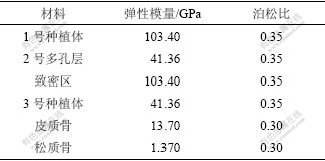
Ӧ��CAD ����Pro/Engineer2001�ڼ�����Ͻ���������ֲ������������ǿ顣�Ե�һǰĥ���ĺ��������Ϊ�����棬������н�Զ�е������γ���ά�����ǹǿ顣�ùǿ���нϺ��Ƥ�ʹ��Լ��ܶȽϸߵ����ʹ�(ģ��Lekholm ��Zarb �Ƿ����B/2��)�������ն���Ƥ�ʹǺ��Ϊ2.0 mm���ǿ�ļ��β���Ϊ����35 mm�������14 mm����Զ�г�20 mm�����ʹǵĽ�Զ��δ��Ƥ�ʹǰ��ơ�ģ��һԲ������ֲ�壬����ֲ��ĵ���ģ���ͽṹ�ص㣬����ȫ�����͡�����͵���ģ���ͺʹ�Ƥ�������ܶ����ʹ������������о������3������ֲ�壬ֱ�����߾�Ϊ4.1 mm��12 mm��ͬʱģ���Ϊ5 mm�Ļ�̨����̨����ֲ���Ϊһ��ͬ�����塣
Ӧ��Ansys Workbench 10.0 ������������Ԫ����������Ansys��Pro/E��ר�ýӿڣ���ʵ��ģ�͵���Ansys����3Dʵ�嵥Ԫ(solid45)������ͬ�����ܶȵ��������֣������ǿ�����ֲ�����ά����Ԫģ�ͣ�ͨ����������Ļ��֣����Ƶ�Ԫ�Ĵ�С�����Ե�Ԫ�����Զ��Ż���ģ����ͼ1��ͼ2��ʾ����Ԫ����ڵ������£���ֲ��ģ����2 498�ڵ㡢11 501��Ԫ����ģ����6 425�ڵ㡢33 382��Ԫ��
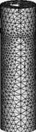
ͼ1 ��ֲ������Ԫģ��
Fig.1 Finite element models of implant
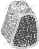
ͼ2 ������Ԫģ��
Fig.2 Finite element models of partial mandibular bone
��ģ���еĸ��ֲ��Ϻ���֯����Ϊ���������ȡ�����ͬ�Ե�С�����ߵ��Բ��ϣ���ֲ�������֯���������Ϊ100%�Ĺ��Խ�ϡ���Ƥ�ʹ�ģ�͵IJ��漰���ʹ�ģ�͵IJ���͵�����ȫԼ���������˱߽�������չ����Ӧ�ڵ��ϡ�����ֲ��������϶�ģ��ʩ����ͬ��ȫԼ���������Ƹ���������ֲ������ɶȡ�1������ֲ��Ϊȫ�����ͣ�2��Ϊ��Ƥ�������ܶ����ʹ����������о�����ͣ�3��Ϊ����͵���ģ���͡�������ֲ��ṹ���ضԹǽ���Ӧ��Ӱ��ʱ�����������ѵĵ���ģ��103.4 GPa��2�Ŷ�ײ��3�ŵĵ���ģ��Ϊ������ģ����40%��������ֲ�嵯��ģ����2�ź�3����ֲ��ǽ���Ӧ��Ӱ��ʱ������4�ֵ���ģ���ֱ�Ϊ������ģ����20%��40%��60%��80%�����ֲ��ϵ���ѧ�������1��ʾ��
��1 ģ��ĸ�������ֲ�����ѧ����
Table 1 Mechanical properties of used titanium implants
2 ģ�������
2.1 ��ͬ�ṹ������ֲ��ǽ���Ӧ���ֲ��Ƚ�
ģ��������ҧ���˶����Խӽ���������ҧ����������ֲ���-�ǿ�ģ�ͼ��أ��ֱ���������150 N �ʹӼ�����45?����25 N��
ȡVon MisesӦ����Ϊ����Ӧ��ˮƽ����Ҫָ�ꡣ�ȽϷ���3������ֲ������Ӧ���ֲ��Լ���ģ�ǽ�����Ӧ���ֲ�״����
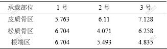
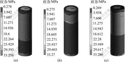
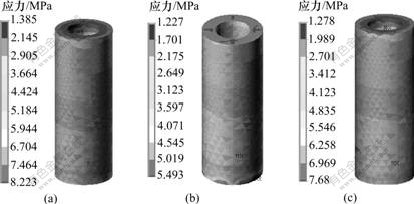
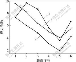
����ͬ�غ��£�3������ֲ��ǽ������Von-MiseseӦ����Ƥ�ʹǡ����ʹǺ���������ͬ����Ƥ�ʹ�����3����ֲ���Von-MiseseӦ����ߣ�Ϊ7.128 MPa���������ʹ�����������1����ֲ���Von-MiseseӦ����ߣ�Ϊ6.704 MPa(����2)���ӽ���Ӧ���ֲ���ͼ�ɿ�����3������ֲ�徱��Ƥ�ʹǾ�Ϊ��Ӧ����(��ͼ3)��Ƥ�ʹ������ʹǽ�������������Ϊ��Ӧ������1����3����ֲ�������ʹ����ĸ�Ӧ���������2����ֲ��ĸ�Ӧ��(��ͼ4)�������ʹ�����2����ֲ���Von-MiseseӦ������1����3����ֲ���Ӧ�����ҳʾ��ȵݼ�����(��ͼ5)��
��2 3�ֲ�ͬ�ṹ����ֲ��ǽ������Von-MiseseӦ��
Table 2 Maximum Von Mises stresses in implant-bone interface of three different structure titanium implants p/MPa
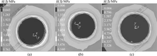
(a)1��; (b) 2��; (c) 3��
ͼ3 3������ֲ��Ƥ�ʹ���Von-MiseseӦ���ֲ���ͼ
Fig.3 Stress distribution in cortical bone area of three titanium implants
(a) 1��; (b) 2��; (c) 3��
ͼ4 3������ֲ������ǽ���Von-MiseseӦ���ֲ���ͼ
Fig.4 Stress distribution in axial bone-implant interface of three titanium implants
(a) 1��; (b) 2��; (c) 3��
ͼ5 3������ֲ�������ʹ����ǽ����Ӧ���ֲ���ͼ
Fig.5 Stress distribution in cancellous bone-implant interface of three titanium implants
2.2 ��ͬ�ṹ��ֲ������ǽ���Von-MiseseӦ���ֲ��ıȽ�
������ֲ��������λ�ù�ϵ����Ƥ���������ʸ��˲�����ֲ�峤�᷽�����6�����棬����1 Ϊ����������4 Ϊ�������в�������6 Ϊ���˲�����������ػ���������������ϼ����£�3����ֲ��Ľ���Ӧ���ֲ�״�����£�2����ֲ���Ƥ���������ʹ�������Ӧ���ʾ��Ƚ������ƣ������Ӧ����1�ź�3����ֲ��ĵͣ�1����ֲ�������ʹ����Ľ���Ӧ����2�ź�3�ŵĸߣ�3����ֲ����Ƥ��������Ӧ�����������ڸ�������3����ֲ�����Ӧ������������1����ֲ��Ӧ����������(��ͼ6��ͼ7)��
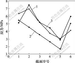
1��1��; 2��2��; 3��3��
ͼ6 ���������3�ֲ�ͬ��ֲ��ǽ���Ӧ���ֲ�ͼ
Fig.6 Stress distribution in bone-implant interface of three different titanium implants in an axial loading
1��1��; 2��2��; 3��3��
ͼ7 ���ϼ�����3�ֲ�ͬ��ֲ����ѹ�����Ӧ���ֲ�ͼ
Fig.7 Stress distribution in oppressed flank of three different titanium implants in united loading
2.3 ��ͬ����ģ����2����3����ֲ��ǽ���Ӧ���ֲ��ıȽ�
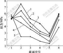
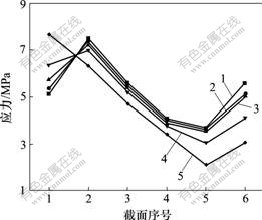
����4�ֵ���ģ���ֱ�Ϊ������ģ����20%��40%��60%��80%������ͬ�غ��£��о�����ģ����2����3����ֲ��ǽ���Ӧ���ֲ���Ӱ�졣�о����֣��ڸ����£�2����ֲ��ǽ���Ӧ���浯��ģ���Ľ��Ͷ���С�������Ա�ȫ��������ֲ��ĵͣ�����ײ��ģ��Ϊ�����ѵ�40%������ʱ�����ʹ�������Ӧ����������(��ͼ8)����3����ֲ��ǽ���Ӧ���浯��ģ�����ͣ�Ƥ�ʹ�������Ӧ�����ӣ������ʹ�������Ӧ�����Ͳ�����(��ͼ9)��
1��ȫ���ܣ�2��80%ģ����3��60%ģ����
4��40%ģ����5��20%ģ��
ͼ8 ��������²�ͬ����ģ����2����ֲ��ǽ���Ӧ���ֲ�ͼ
Fig.8 Stress distribution in bone-implant interface of different elastic modulus No.2 titanium implants in an axial loading
1��ȫ���ܣ�2��80%ģ����3��60%ģ����
4��40%ģ����5��20%ģ��
ͼ9 ��������²�ͬ����ģ����3����ֲ��ǽ���Ӧ���ֲ�ͼ
Fig.9 Stress distribution in bone-implant interface of different elastic modulus No.3 titanium implants in an axial loading
3 ����������
��ֲ����Ҫ��������Χ����֯��ֱ�ӽ����ʹ���ܣ��ǽ�Ͻ���Ŀɿ��Ժ��ȶ��Ծ�����ֲ�����ڳɹ��ʡ�Ҫȷ����ֲ�峤���ȶ�����ʹ���ܣ�һ���澡�������غɣ����ⳬ�������������ȣ����غ����½���������ջ���ֲ��Ӧ��ƣ��ʧ�ܣ������غɿ���������Թ�ή������ɥʧ[5-6]����һ���棬Ҫ������ֲ��ı����������������ǵĽӴ���������ǽ���Ӧ����Ϊ������ֲ�������֯�Ľ��ǿ�Ⱥ����������Ըı���ֲ����״���������������ϸ��ֳɹ����ӵȴ�ʩ����������ֲ�������֯�ĽӴ�����ǽ��ǿ�ȣ�������ֲ���������ѧ������
��ҽѧ��������ѧ�Ƕȿ������������Ӳ�Ȳ���̫�ߣ����⽵���¹��γ����踺�ɣ���ɹ�Ӧ�����գ����ڲ���Ӧ���ڳ��ز�λ��Ҫ���㹻�Ŀ�ƣ��ǿ�ȣ�ֲ��������ϵĵ���ģ��ԽС��Խ�ӽ��ǵĵ���ģ���������ڳ���Ӧ��ʱ����Ӧ�������ɵ����λ��ԽС���Ӷ���С�����ɶ�������Ӧ��������ɵĹ����պ��˻�����Ȼ�ǵ�ǿ�ȷ�ΧΪ3~20 MPa��Ƥ�ʹǵĵ���ģ��Ϊ10~18 GPa�����ʹ�Ϊ1.3~4 GPa������ģ���Կ�϶���У���ʹ25%�Ŀ�϶��Ҳ��������ģ���½�50%[7-9]�����ж��ؽṹ�Ķ��ֲ������ܼ�������ֲ��������������[10-12]�������ڳɹ�ϸ����ճ��������ϸ������ʳ�����Ӫ�����������룬��ʹ�����dz����϶��ʵ������̶���ͨ���ı��϶���������������ϵ��ܶȡ�ǿ�Ⱥ͵���ģ�����ﵽ�뱻�滻Ӳ��֯��ƥ�����ѧ���ܣ�ʵ�ֲ��ϵ���ѧ�����ԣ���ṹΪ�������Ϳ���ṩ֧�ܣ��ٽ�������[13-15]��
����ģ��������������ͬ�غ��£�ȫ�����ͺ�����͵���ģ������ֲ������Ӧ���ֲ����ɴ�����ͬ���������£���ֲ�徱����ΧƤ�ʹ����Ľ���Ӧ�������ϳ����������ƣ���Ƥ�ʹ������ʹǵĽ��粿����һ�����Ե��½����̣������ʹǽ�������ֲ�峤��ķֲ�����Ϊ�ݼ����ڸ�����Ӧ�����ӣ���������ѧ�����ó��ķֲ�����һ��[3-4, 8]������Ƥ�������ܶ����ʹ����������о��������ֲ��Ľ���Ӧ����Ƥ�ʹǺ����ʹ���������Ϊ�ݼ���������Ӧ�����ӣ������ʹ�������ֲ��ǽ���Ӧ������ȫ�����ͺ�����͵���ģ������ֲ��Ӧ������˵������ƴ�Ƥ�������ܶ����ʹ����������о����Ϊ�ṹ���Ե���ֲ������Ч�ؽ�����ֲ��ǽ���Ӧ���������ڹǽ���Ӧ��ת������Χ����֯��
4 �� ��
a. ����ͬ�غ��£�3������ֲ�徱��Ƥ�ʹǾ�Ϊ��Ӧ������3����ֲ���Ӧ����ߣ�Ϊ7.128 MPa�������ʹ���2����ֲ���Ӧ����1����3����ֲ��ĵͣ��ҳʾ��ȵݼ����ơ�
b. �ڼ��������£�2����ֲ�����ŵ���ģ���Ľ��ͣ��ǽ���Ӧ�����ͣ������Ա�1����ֲ��ĵͣ�����ײ�ĵ���ģ��Ϊ�����ѵ�40%������ʱ���ǽ���Ӧ�����Խ��ͣ� 3����ֲ�����ŵ���ģ�����ͣ�Ƥ�ʹ����ǽ���Ӧ�����ӣ������ʹ����ǽ���Ӧ�����Ͳ����ԡ�
c. ����ֲ��Ľṹ�͵���ģ����Ӱ��ǽ���Ӧ���ֲ�����Ƥ�������ܶ����ʹ����������о����������ֲ�������ڽ���Ӧ��ת�Ƶ���Χ����֯�����Ͷ�ײ�ĵ���ģ��������Ч�ؽ��ǽ���Ӧ����
�ο����ף�
[1] Van Osterwyck H, Duyck J, Vander S, et al. The influence of bone mechanical properties and implant fixation upon bone loading around oral implants[J]. Clin Oral Implants Res, 1998, 9(6): 407-412.
[2] Geng J, Tan K B, Liu G R. Application of finite element analysis in implant dentistry: A review of the literature[J]. J Prosthet Dent, 2001, 85(6): 585-598.
[3] Iplikcioglu H, Akca K. Comparative evaluation of the effect of diameter, length and number of implants supporting three-unit fixed partial prostheses on stress distribution in the bone[J]. J Dent, 2002, 30(1): 41-46.
[4] ����, ������, ���Ľ�, ��. Ӱ���˹���ֲ��-�ǽ���Ӧ���ֲ��������صĶ�Ԫ�ع����[J]. ҽ��������ѧ, 2000, 15(4): 216-221.
ZOU Jing-cai, LIU Bao-lin, TANG Wen-jie, et al. Multivariate step regression analysis of influencing factors on the stress distribution patients at the bone interface around dental implants[J]. Journal of Medical Biomechanics, 2000, 15(4): 216-221.
[5] Pilliar R M, Deporter D A, Watson P A, et al. Dental implant design effect on bone remodeling[J]. J Biomed Mater Res, 1991, 25(4): 467-483.
[6] Vaillancourt H, Pillar R M, McCammond D. Factors affecting cortical bone loss with dental implants partially covered with a porous coating: A finite element analysis[J]. Int J Oral Maxillofac Implants, 1996, 11: 351-359.
[7] Sato Y, Wadamoto M, Tsuga K, et al. The effectiveness of element downsizing on a three-dimensional finite element model of bone trabeculae in implant biomechanics[J]. J Oral Rehabil, 1999, 26(4): 288-291.
[8] Sahin S, Cehreli M C, Yalcn E. The influence of functional forces on the biomechanics of implant-supported prostheses��A review[J]. J Dent, 2002, 30(8): 271-282.
[9] Becker B S, Bolton J D, Youseffi M. Production of porous sintered Co-Cr-Mo Alloys for possible surgical implant applications[J]. Powder metallurgy, 1995, 38(3): 201-208.
[10] St-Pierre J P, Gauthier M, Lefebvre L P, et al. Three-dimensional growth of differentiating MC3T3-E1 pre-osteoblasts on porous titanium scaffolds[J]. Biomaterials, 2005, 26(35): 7319-7328.
[11] Otsukia B, Takemotoa M, Fujibayashia S, et al. Pore throat size and connectivity determine bone and tissue ingrowth into porous implants: Three-dimensional micro-CT based structural analyses of porous bioactive titanium implants[J]. Biomaterials, 2006, 27(35): 5892-5900.
[12] Takemoto M, Fujibayashi S, Neo M, et al. Mechanical properties and osteoconductivity of��porous bioactive titanium[J]. Biomaterials, 2005, 26(30): 6014-6023.
[13] Oguz K, Emir Y, Fehmi E. Static, dynamic and fatigue behaviors of dental implant using finite element method[J]. Advances in Engineering Software, 2006, 37(10): 649-658.
[14] �� ��, �����, ������.����ģ���ͳ�ʼӦ������ֲ��ǽ���Ӧ���ֲ�Ӱ�����ά����Ԫ����[J]. �й���ǻ��ֲѧ��־, 2006, 11(2): 55-58.
ZHAO Feng, HAN Yan-feng, HU Jiang-feng. Three-dimensional finite element method analysis of relation of implant elastic modulus and initial stress and bone-implant surface stress distribution[J]. Chinese Journal of Oral Implantology, 2006, 11(2): 55-58.
[15] �� ��, �� ��, ������.��������ֲ�嵯��ģ����ǽ���Ӧ���ֲ���ϵ����ά����Ԫ����[J].�ٴ���ǻҽѧ��־, 2006, 22(11): 667-670.
GAO Bo, ZHAO Feng, LIU Zhen-xia. Three-dimensional finite element method analysis of relation of screw implant elastic modulus and bone-implant surface stress distribution[J]. Journal of Clinical Stomatology, 2006, 22(11): 667-670.
�ո����ڣ�2008-09-10�������ڣ�2008-11-20
������Ŀ��������Ȼ��ѧ����������Ŀ(35770576)����863���ƻ��²��ϼ�������ר��������Ŀ(2007AA03Z114)������ʡ��Ȼ��ѧ����������Ŀ(2007JJ5109)
ͨ�����ߣ�������(1967-)���У����������ˣ���ʿ�������ڣ�������������ֲ�忪�����ٴ��о����绰��0731-8618554��E-mail: chen0313@xy3yy.com
" target="blank">[15] �� ��, �� ��, ������.��������ֲ�嵯��ģ����ǽ���Ӧ���ֲ���ϵ����ά����Ԫ����[J].�ٴ���ǻҽѧ��־, 2006, 22(11): 667-670.GAO Bo, ZHAO Feng, LIU Zhen-xia. Three-dimensional finite element method analysis of relation of screw implant elastic modulus and bone-implant surface stress distribution[J]. Journal of Clinical Stomatology, 2006, 22(11): 667-670.
