

Effect of porous structure on mechanical properties of
C/PLA/nano-HA composites scaffold
LIAO Xiao-ling(������)1,2, XU Wen-feng(���ķ�)3, WANG Yuan-liang(��Զ��)1,
JIA Bi(�� ��) 2, ZHOU Guan-yu(�ܹ��)2
1. College of Bioengineering, Chongqing University, Chongqing 400044, China;
2. College of Metallurgical and Materials Engineering, Chongqing University of Science and Technology,
Chongqing 401331, China;
3. College of Chemistry and Environment Engineering, Chongqing University of Arts and Sciences,
Chongqing 402160, China
Received 10 August 2009; accepted 15 September 2009
Abstract:
Nonporous and porous C/PLA/nano-HA composites were fabricated by the process of solvent blending and freeze-drying technique, and the effect of porous structure on the mechanical properties of C/PLA/nano-HA composites scaffold was investigated and analyzed. The results show that the effects of porous structure on the bending strength, modulus and curves of stress and strain were obvious. Compared with nonporous sample, the curves of stress and strain of porous sample show more rough, and alternative phenomenon of stress increase and stress relaxation appears. It is strongly suggested that the fracture model of C/PLA/nano-HA composites scaffold transforms from the local to global load due to the porous structure.
Key words:
porous structure; mechanical properties; C/PLA/nano-HA composite;
1 Introduction
Hard tissue defect is a serious problem that affects the health of people due to trauma, tumor, inflammation and social aging. With the development of material science, biomedicine and bionics, the tissue engineering approach to repair or replace has been demonstrated successful in tissue regeneration, in which the tissue engineering material plays an important role in moulding regenerated tissue and organs, and provides an environment of nutrition and gas exchange and a spaces for cell proliferation, so porous structure was required for tissue engineering scaffold[1- 4].
Today, various types of porous tissue engineering materials have been developed[5-10], of which polylatide (PLA), hydroxyapatite(HA) and Chitosan(CS) were most widely employed to build bone tissue engineering scaffolds[11]. But the single PLA, HA, or CS cannot meet the requirements of mechanical strength for massive bone. In order to solve this problem, the inorganics/organics composites are the inevitable way, especially the fiber reinforced composites. In this work, the carbon fiber (CF) reinforced PLA/nano-HA biomaterial composites (C/PLA/nano-HA) were prepared by the process of solvent blending and freeze-drying technique, in which the CF was used as the reinforcement to improve mechanical properties, and the advantages of PLA/nano-HA were retained. So, the composites will be a bioactive, absorbable, degradable and high mechanical internal fracture fixation materials.
As repair or replacement materials of hard tissue defect, the potential application of C/PLA/CS composites is inevitable to involve mechanical loading, and the porous structure is also necessary, so we have to pay attention to the effect of porous structure on mechanical properties of C/PLA/CS composites[12]. However, there is just deficiency in the effect of porous structure on mechanical properties for these repair or replacement materials, and the fracture behavior is not also clear. Therefore, in this work, the research is focused on the effect of porous structure on the bending strength, modulus and curves of stress and strain, finally the mechanism is discussed.
2 Experimental
2.1 Materials
2.1.1 Preparation of materials
Poly(l-lactide) (PLA; Mn=200 kDa and Mw=350 kDa) was synthesized, provided and purchased from College of Bioengineering, Chongqing University (Chongqing, China). Hydroxyapatite (biochemical reagent) was obtained from Sinopharm Chemical Reagent in Beijing Co. Ltd(Beijing, China). Carbon fiber(T300, d=7-8 ��m, ��=1.78 g/cm3) was provided by Toray Co. Ltd (Japan).
2.1.2 Preparation of composites
In this work, the C/PLA/nano-HA composites were fabricated by the process of solvent blending and freeze-drying technique. The mass ratio of PLA to nano-HA was 85?15, and the volume fraction of CF was 10%. The PLA was dissolved with ethyl acetate, and then added with nano-HA, mixed using ultrasonic for 4 h.
The preparation of nonporous composites was as follows. The dispersed CF was used to infiltrate adequately the PLA/nano-HA mixture to form the pro infiltration banding, and then these infiltration bandings were fabricated to be rectangular nonporous composites with 6 mm in thickness in the laid-up direction and 14 mm in width and 40 mm in length by using compression molding.
The preparation of porous composites was as follows. The CF/PLA/nano-HA mixture was rapidly put into freeze-drying equipment to make pore, and then was processed into rectangular porous composites with size of 6 mm��12 mm��40 mm.
2.2 Mechanical test
The dimensions of the specimens of the C/PLA/nano-HA composites used for three point bending test were 3 mm��6 mm��40 mm. And the five specimens were tested for each condition. All the tests were performed using a servo hydraulic model SANS2CMT4204 testing machine with an automated data-acquisition system.
2.3 Characterization of porous composites
After vacuum drying at 35 ��, the samples were sprayed with gold using ion sputtering technique, and the surface morphology and microstructure were observed using scanning electron microscopy (SEM Hitachi Model S3400N).
The porosity of the composites was measured by pycnometer method using ethanol as discharge liquor. The porosity was calculated by the following formula:
Porosity=(volume of pores/total volume of materials) ��100%
3 Results and discussion
3.1 Porous structure and mechanical properties
Fig.1 shows the relationship between bending strength, modulus and the porosity of C/PLA/nano-HA composites. It is clear that the bending strength and modulus decrease with increasing porosity, which is explained by the fact that the porous structure leads to the decrease of carrying capacity of materials. This typical behavior has been reported by many researchers, and has been vaguely explained that the densification is assumed to cause relief in the stress concentrations in the matrix due to the minimization of undesirable matrix defects[13-15]. So, the porous structure obviously increases more defects in the composites, and more pores mean more defects.
The porous microstructure of the C/PLA/nano-HA composites is shown in Fig.2. From the arrows in Fig.2,the pores cavitate obviously the interface of CF and matrix, which mainly weakens the interface strength and decreases the mechanical properties of the composites.

Fig.1 Effect of porosity on flexural strength and modulus of C/PLA/nano- HA composites
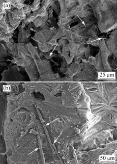
Fig.2 Effect of porosity on structure of C/PLA/nano-HA composites
3.2 Porous structure and curves of stress and strain
Fig.3 shows the curves of stress and strain of nonporous and porous composites with various porosities. It is clear that the fracture behavior of the porous sample is much different from that of the nonporous one. Compared with the nonporous sample, the curves of stress and strain of porous samples show more rough, and alternative phenomenon of stress increase and stress relaxation appears, as shown in Fig.3.
These results demonstrate that porous structure gives rise to the nonlinear behaviors. This is mainly attributed to porous matrix and weak interfaces between the fibers and matrix. The fracture patterns of the C/PLA/nano-HA composites are considered as follows: the fracture model transforms from the local to global load.
The local load-sharing regime (LLS) appears in composites with strong interfaces between the fibers and matrix[6]. This process continues and thus results in a catastrophic ultimate fracture as shown in Fig.3(a). On the other hand, the global load sharing regime (GLS) appears in composites with weak interfaces. In this regime, with the beginning of loading, the pore walls of porous materials will bend, and linear elasticity appears. But when loading reaches the critical stress, the pores start to collapse, and the characteristic of brittle fracture appears due to the brittle fracture of pore walls. When the composites are in high strain, the pores fully collapse, which leads to the structure of pore walls producing a series of deformation, such as touching with each other, pressing together, broken splinter accumulation, and finally form the densification of porous materials. This can cause the increase of stress and strain. This process is then repeated. The ultimate elongation in the GLS regime should be larger than that in the LLS regime. The stress�Cstrain curve becomes nonlinear when multiple-deformation of the porous structure begins, and alternative phenomenon of stress increase and stress relaxation appears.
4 Conclusions
1) The porous structure decreased the bending strength and modulus, and the fracture behavior of the porous sample was much different from that of the nonporous one.
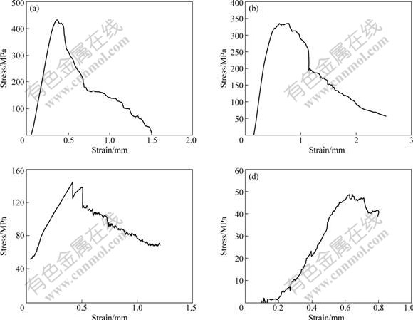
Fig.3 Curves of stress and strain of C/PLA/nano-HA composites: (a) Nonporous composites; (b) Porous composite with porosity of 20%; (c) Porous composite with porosity of 60%; (d) Porous composite with porosity of 80%
2) Compared with nonporous sample, the curves of stress and strain showed more rough, and alternative phenomenon of stress increase and stress relaxation appeared.
3) It is strongly suggested that the fracture model of C/PLA/nano-HA composites scaffold transforms from the local to global load due to the porous structure.
References
[1] HO M H, KUO P Y, HSIEH H J, WINN S R. Preparation of porous scaffolds by using freeze-extraction and freeze-gelation methods [J]. Biomaterials, 2004, 25: 1129-1135.
[2] WILLIAMSA J M, ADEWUNMI A, FLANAGAN C L. Bone tissue engineering using poly scaffolds fabricated via selectivelaser sintering [J]. Biomaterials, 2005, 26: 4817-4827.
[3] KIM K, YU M, ZONG X H, CHIU J, FANG D F, SEO Y S, HSIAO B S, CHU B, HADJIARGYROU M. Control of degradation rate and hydrophilicity in electrospun non-woven poly(D,L-lactidc) nanofiber scaffolds for biomedical applications [J]. Biomaterials, 2003, 24(27): 49-77.
[4] NAM Y S, PARK T G. Porous biodegradable polymeric scaffolds prepared by thermally induced phase separation [J]. J Biomed Mater Res, 1999, 47(1): 8-12.
[5] BORDEN M, EL-AMIN S F, ATTAWIA M. Structural and human cellular assessment of a novel microsphere-based tissue engineered scaffold for bone repair [J]. Biomaterials, 2003, 24(4): 597-609.
[6] NAM Y S, YOON J J, PARK T G. A novel fabrication method of macroporous biodegradable polymer scaffolds using gas foaming salt as a porogen additive [J]. J Biomed Mater Res, 2000, 53(1): 1-7.
[7] HARRIS L D, KIM B S, MOONEY D J. Open pore biodegradable matrices formed with gas foaming [J]. J Biomed Mater Res, 1998, 42(3): 396-340.
[8] ESPINOSA H D, RAISER G, CLIFTON R J, ORTIZ M. Experimental observations and numerical modeling of inelasticity in dynamically loaded ceramics [J]. Journal of Hard Materials, 1992, 3(3/4): 285-313.
[9] WU Lin-bo, JING Dian-ying, DING Jian-dong. A ��room temperature�� injection molding/particulate leaching approach for fabrication of biodegradable three dimensional porous scaffolds [J]. Biomaterials, 2006, 27(2): 185-190.
[10] YANG K F, LEONG Z D, CHUA C K. The design of scaffolds for use in tissue engineering (Part II): Rapid prototyping techniques [J]. Tissue Eng, 2002, 8(1): 12-15.
[11] YAO J, RADIN S. The effect of bioactive glass content on synthesis and bioactivity of composite poly (lactic-co-glycolicacid)/bioactive glass substrate for tissue engineering [J]. Biomaterials, 2005, 26(14): 1935-1943.
[12] TABOAS J M, MADDOX R D, HOLLISTER S J. Indirect solid free form fabrication of local and global porous biomimetic and composite 3D polymer-ceramic scaffolds [J]. Biomaterials, 2003, 24: 181-194.
[13] HATTA H, SUZUKI K, SHIGEI T, et al. Strength improvement by densification of C/C composites [J]. Carbon, 2001, 39: 83-90.
[14] ELI YOON J J, PARK T G. Degradation behaviors of biodegradable macroporous scaffolds prepared by gas foaming of efferves cent salts [J]. J Biomed Mater Res, 2001, 55(3): 401-408.
[15] ZAVATTIERI P D, ESPINOSA H D. An examination of the competition between bulk behavior and interfacial behavior of ceramics subjected to dynamic pressure-shear loading [J]. Journal of the Mechanics and Physics of Solids, 2003, 51: 607-635.
(Edited by YUAN Sai-qian)
Foundation item: Project(30870609) supported by the National Natural Science Foundation of China; Projects(KJ081205; KJ091213) supported by the Natural Science Foundation of Chongqing Education Committee, China
Corresponding author: LIAO Xiao-ling, Tel: +86-23-65023701; E-mail: zxc_228@163.com
