
����������˾��д�С���γ���ػ���mpsA�ڲ�ͬ��Դ�̼��µIJ������
�����ǣ����ı��� ӱ���亣�ޣ������
(���ϴ�ѧ ��Դ�ӹ������﹤��ѧԺ������ ��ɳ��410083)
ժ Ҫ��
ժ Ҫ��Ϊ�о�����������˾�(Acidithiobacillus ferrooxidans)�ڰ����γɵĵ������ܵĴ��Կ�������ػ�������������˾�������ATCC23270��ȫ�������������Ϣѧ���з�������ATCC23270��ȫ�������ϲ���������ϸ����mpsA�����ͬԴ����ORF1622����������б��ؽṹ���������бȶ��Լ�������ͬԴ�Է��������÷�ת¼PCR������ת¼ˮƽ�о�mpsA�����������������·ֱ���20 mmol/L FeCl3��FeSO4��7H2O�̼�ʱ�IJ����������֤�����ڴ�С���γɹ����е����á��о����������ORF1622����ĵ�����PRK05724�ṹ����mpsA������ͬ��Ϊ48%����acetyl-CoA carboxylase carboxyltransferase subunit alphaͬԴ������������˾��е�mpsA������ת¼����ı�����������ֱ�ӹ�ϵ����������������˾������������������ɴ�С�壬��ˣ���������������˾��д�С����γ���ء�
�ؼ��ʣ�
����������˾���mpsA������������Ϣѧ��������ת¼PCR��
��ͼ����ţ�Q786 ���ױ�־�룺A ���±�ţ�1672-7207(2009)06-1471-05
Real-time PCR analysis of different Fe ion shock responses of mpsA gene in Acidithiobacillus ferrooxidans ATCC 23270
LIU Xin-xing, LIU Wen-bin, YAN Ying, WU Hai-yan, QIU Guan-zhou
(School of Minerals Processing and Bioengineering, Central South University, Changsha 410083, China)
Abstract: The whole genome of the type strain Acidithiobacillus ferrooxidans ATCC 23270 and the conserved domain and amino acid sequences as well as the protein identity of the target gene were analyzed. To obtain further knowledge of the magnetosomes formation mechanisms of Acidithiobacillus ferrooxidans in response to different kinds of Fe ions, temporal gene expression profiles were examined in cells subjected to 20 mmol/L FeCl3 and FeSO4��7H2O shock by using reserved transcript PCR. The results indicate that ORF 1622 of the ATCC 23270 is homologous with mpsA gene in magnetotactic bacteria and the expressions of ORF 1622 is related to Fe2+. According to the fact that the Acidithiobacillus ferrooxidans is able to synthesize intracellular magnetosomes when it grows on FeSO4, it can be supposed that the mpsA gene is related with the magnetosome formation in Acidithiobacillus ferrooxidans.
Key words: Acidithiobacillus ferrooxidans; mpsA gene; bioinformatics analysis; RT-PCR
����������˾�(Acidithiobacillus ferrooxidans)��Temple��[1]��20����50��������ֲ������ġ���������ұ���е���Ҫϸ��֮һ���������û�����ȿ����е���������Ӷ�ʹ����ֽ⣬�ͷų����������еĹ��������ˣ��㷺���ڽ������ֽ����������ǵ�[2]������������˾��з����˴��Կ�������С��ϸ����ѧ���ȸߣ��ᾧ��������С���ȣ������Ի���ϸ�����������õ����������ԣ����ԣ�����һ��������������ײ���[3-6]��Ŀǰ��Grunberg��[7-13]�ӵ����ʺͻ���Ƕ��о��˴�С����γɻ�����Matsunaga��[14]����SDS-PAGE������Magnetospirillum sp. AMB-1����õ�mpsA�������ĵ��ס��û������Ϊ1����317������IJ���Nĩ��ϸ�������ź����еĵ����ʣ�ͬԴ��������mpsA������˾�����-CoA�Ȼ�ø�Ħ��ǻ�����52%��ͬԴ�ԣ����Ҿ���CoA�Ľ������û��ֱ��֤�ݱ�����С��Ĥ��ϸ����Ĥ�����������ڴ�С��Ĥ��ϸ����Ĥ��ɴ�����ͬ������Pfanner��[15]�ı������������Ĥ��ADP-���ǻ����������ӵ�����������Ĥ���ݣ�����֬��-CoA�ܹ��̼�ϸ����߶����ݳ�ѿ�γ�С���ݣ���ˣ�Matsunaga��[14]�Ʋ�MpsA��������CoA��Ϊ�Ȼ�����Ӷ�ת��������������ӣ�ͨ������������ϸ���������γɴ�С��Ĥ����ϸ���ڵ�Fe3O4������
���ڶ�����������˾��д�С���γ���ػ�����о����٣���ˣ���������ͨ��������Ϣѧ������������������˾�ATCC23270������Ѱ�ҵõ���AMB-1������mpsA�����ͬԴ�������÷�ת¼PCR�����о��û���������������������Fe2+��Fe3+�̼��IJ�����̽������������˾��γɴ�С����ػ����Ա�Ϊ�о�����������˾��д�С���γɵ���ػ������»�����
1 ʵ������뷽��
1.1 �� ��
1.1.1 ������������
A.ferrooxidans ATCC 23270���������ֱ�������(American Type Culture Collection��ATCC)������
��������ģʽ���ֱ�������ATCC medium 2039�������䷽��
��Դ������Ũ��Ϊ10 g/L����ۣ���Դ�ֱ�ΪFeSO4��FeCl3�������Լ���Ϊ��������
1.1.2 �����Լ�
RNA��ȡ�Լ�ΪTrizol(Invitrogen)��RNA�����Լ���ΪSV Total RNA Isolation System(Promega)��RNA��ת¼�Լ�ΪSuperScriptTM��ת¼ø(Invitrogen)���������(Invitrogen)��
1.2 ʵ�鷽��
1.2.1 ����������˾���mpsA�����������Ϣѧ ����
A.ferrooxidans ATCC 23270ȫ���������м��䲿��ע�ʹ�The institute for genome research(tigr)����ã�����tigr���������������BLASTѰ��A.ferrooxidans ATCC 23270��������ϸ����mpsA�����ͬԴ������Gene Runner�����������Ŀ�����Ŀ����Ķ���(ORF)������Ŀ�군�ױ��ؽṹ�������Ŀ�군��������ϸ����ͬԴ���װ��������бȶԽ��ͨ��Gene Doc������á���National Center for Biotechnology Information(NCBI)���������е�ͬԴ�Է�����
1.2.2 ����������˾���mpsA������������
����BlastN�ȶԵõ�ͬԴ�������У�����Primer 5.0������ƻ�����������mpsA1 5��-GGCTATTAT- CGGCGGTCTGGC-3���mpsA2 5��-GCTCCGGGCG- TATCAATGAAC-3�䡣��������ص���Ϊ201bp��
1.2.3 ATCC23270�����������������߲ⶨ
ȡ������ATCC23270���ֽ���2 L�������У�����pH=2.0����1%(��������)����Ϊ��Դ����ת��Ϊ180 r/min��ҡ�����������¶�Ϊ30 �档��Ѫ��������ڹ�ѧ������ֱ�ӹ۲������Һ��ϸ��Ũ�ȣ���δ���ر�˵��������ʵ����ظ�3�Ρ�
1.2.4 ��ͬ��Դ������RNA����ȡ��������ת¼
��ϸ�������������ں������õ�ϸ�������ֳ�3�飬���У���1�͵�2��ֱ����FeCl3��FeSO4��7H2O(��Ũ��Ϊ20 mmol/L)�����������½��д̼�����3���������̼�������ϸ����Ϊ�հ��գ��̼�1 h��ÿ��ȡ300 mL����Һ(�¶�Ϊ4 ��)���˳�ȥ��ۺ������䶳�����ģ��ռ����壬ʣ������Һ�����������ȶ����ռ����壬�Ա�����������۲��С������γ�������á�����Trizolһ������ȡ��RNA����RNA�����Լ���(Promega)������RNA����NanoDrop���ֹ��ȼ�(NanoDrop Technologies)���RNAŨ�Ⱥʹ��ȡ�����RNA��mRNAΪģ�壬��ת¼�ϳ�cDNA����NanoDrop���ֹ��ȼƲⶨ��Ũ�ȣ���3��cDNA��ƷŨ�Ⱦ�����200 mg/L����-20 ����ر��á���ת¼����Invitrogen��˾ SuperScriptTM��ת¼ø�����������巴Ӧ�����μ��Լ��в����ֲᡣ
1.2.5 ��ת¼PCR��Ӧ���������ʱ��ͬ��Դ�̼��µIJ������
�ֱ���3����Ʒ��cDNAΪģ�壬���ñ�������������������Ƭ�Ρ�PCR����Ϊ����94 ����� 4 min����94 �����20 s����60 ���˻�20 s����72 ������30 s����30��ѭ������72 �����8 min�����ô�����RNA��Ϊģ��Ϊ���Զ��ա�PCR��Ӧ����1%��֬��������Ӿ���鲻ͬ��Դв���¸�����ı��������
1.2.6 ��ͬ���������´�С���γ����
������ȩ�̶��ռ���ľ��壬�����г�����Ƭ(70 nm)������Ƭ������羵��(JEM1230����ѹ80 kV)���۲�Ƚϲ�ͬ��Դ�̼����ȱ�������µ���������ʱ�����ڴ�С�����������
2 ��������
2.1 ATCC23270��AMB-1mpsA������뵰�ױȶԽ��
��AMB-1�����mpsA������ATCC23270��ȫ��������бȶԵõ�ͬԴ�ĺ������У�Ϊtigr��վATCC23270��ȫ�������ϱ��Ϊ��1622�ŵĻ��û���ȫ��951bp������1����316������ĵ����ʡ��Ըû������ĵ����ʽ��б��ؽṹ������������ͼ1��ʾ���ô˵�����AMB-1 mpsA�������ĵ����ʽ���ͬԴ�ԱȽϣ������ʾ��ͬԴ��Ϊ48%(ͼ2)��acetyl-CoA carboxylase carboxyltransferase subunit alphaͬԴ��
2.2 ATCC23270���������������ߵIJⶨ
![]()
ͼ1 ��mpsAͬԴ��ORF1622���ؽṹ�����
Fig.1 Conserved domain analysis of ORF1622
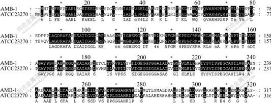
ͼ2 ATCC23270��ORF1622���뵰����AMB-1 mpsA���װ��������бȶԽ��
Fig.2 Amino acid sequences alignment between ORF1622 and mpsA in ATCC23270
Acidithiobacillus ferrooxidans ATCC23270����
��������������ͼ3��ʾ��ͼ3������ATCC23270��������Ϊ��Դʱ�����ڽϳ����ڽ��ֺ��8 d��ȡ��RNA�����о�mpsA�����ڲ�ͬ��Դ�̼��µIJ�����
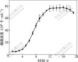
ͼ3 Acidithiobacillus ferrooxidans ATCC23270����������������
Fig.3 Growth curve of Acidithiobacillus ferrooxidans ATCC 23270
2.3 ��RNA����
ϸ����RNA��Ӿ���������ͼ4��ʾ��23S rRNA��16S rRNA����������������������RNA��ȡ���������������Ե�RNase���õ��µĽ��⣬��NanoDrop���ֹ��ȼƼ�⣬�ڲ���Ϊ260 nm��280 nm��������ϵ��OD260��OD280��ֵΪ1.90~2.00����Ʒ�Ĵ��Ƚϸߡ���RNA�Ĵ����������Ծ����ã�mRNA���⡣
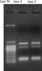
lane MΪmarker��lane 1ΪFe2+�̼�1 h��RNA��lane 2ΪFe3+�̼�1 h��RNA
ͼ4 ��RNA��Ӿͼ
Fig.4 Gel analysis of total RNA
2.4 ��ͬ��Դ�̼�������������˾�mpsA����IJ���������
��ת¼PCR�������֬�ǵ�Ӿͼ����ͼ5��ʾ����ͼ5��֪����ATCC23270��������Ϊ��Դʱ��mpsA�����ڲ�ͬ��Դ�̼��¾��б�����죻mpsA������Fe2+�̼�����cDNA���б����Fe3+�̼��Լ��������̼�ʱ���ޱ��PCR����ԼΪ200bp����������Ƶ�201bp�ص�������һ�£�����PCR����ΪĿ������˵������������˾���mpsA����ı�������Fe2+��صģ��������Լ�Fe3+�ء�
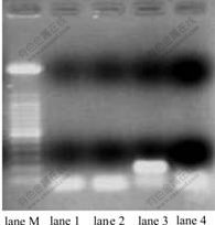
lane1Ϊ�����̼���������lane 2ΪFe3+�̼���lane 3ΪFe2+�̼���lane 4Ϊ���Զ��գ�lane MΪ100bp marker��Ŀ��Ƭ�γ���Ϊ200bp
ͼ5 mpsA����RT-PCR�����Ӿͼ��
Fig.5 RT-PCR analysis of different Fe ion shock response of mpsA gene
2.5 ��ͬ���������´�С���������
��������������ͼ6��ʾ����ͼ6��֪��Acidithiobacillus ferrooxidans ATCC 23270 ��Fe2+�̼�������ʱ�����ɵ������ܵĴ��Կ���(ͼ6(c))�����������̼�������(ͼ6(a))��Fe3+�̼�������ʱ(ͼ6(b)) ��û�е������ܿ����γɡ�
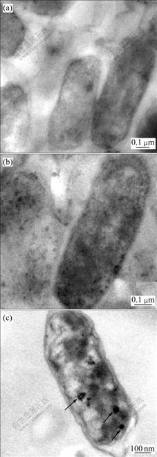
(a) �����̼���(b) Fe3+�̼���(c) Fe2+�̼���
��ͷ��ʾ��ɫ�������ܿ���Ϊ��С��
ͼ6����ͬ��Դ�̼�������������Acidithiobacillus ferrooxidans ATCC 23270������Ƭ��羵��Ƭ
Fig.6 Transmission electron micrograph images of thin-sectioned Acidithiobacillus ferrooxidans ATCC 23270 cells under different Fe ion shocks
3 �� ��
a. �������������˾�ATCC23270��ȫ���������ҵ�������ϸ��Magnetospirillum sp.AMB-1�����д�С��ϳɻ���mpsA��ͬԴ���������������ĵ����бȶԣ������Ըߴ�48%��
b. ATCC23270������mpsA�������������������£���Fe2+�̼����б����Fe3+�̼��Լ��������̼�ʱ���ޱ����˵��mpsA����������������˾��������Ĵ�л��أ�����ATCC23270�д�С����γ���������ض������ʼ��������ص������� һ�¡�
c. ����mpsA����������ϸ����Ϊ��С��ϳɵ���Ҫ���û���������������˾��Ĵ�С���γɹ�����Ҳ�������ش����ã���������С��Ĥ������أ���������÷�ʽ�����ػ������д��ڽ�һ���о���
d. ����������Ϣѧ�������RT-PCR�������ܹ�������������˾����ҵ�����Ŀ������С���γ���صĻ����⽫Ϊ�о�����������˾��д�С����γ��ṩһ����Ч�ֶΣ�Ҳ��Ϊ̽������������˾��д�С���γɻ����Լ�������ػ��ĵ���������»�����
�ο����ף�
[1] Colmer A R, Hinkle. The role of microorganisms in acid mining drainage1a preliminary report[J]. Science, 1947, 106: 253-256.
[2] ������, ���ı�, �� ӱ, ��. ����ƽ���Ӿ����ϸ���·������о�[J]. �й����﹤����־, 2008, 28(3): 79-83.
LIU Xin-xing, LIU Wen-bin, YAN Ying, et al. Isolation of acidithiobacillus ferrooxidans by using solid-plate magnetophoresis[J]. China Biotechnology, 2008, 28(3): 79-83.
[3] Matsunaga T, Nakayama H, Okochi M, et al. Fluorescent detection of cyanobacterial DNA using bacterial magnetic particles on a MAG-microarray[J]. Biotechnology and Bioengineering, 2001, 73(5): 400-405.
[4] Yoza B, Matsumoto M, Matsunaga T. DNA extraction using modified bacterial magnetic particles in the presence of amino silane compound[J]. Journal of Biotechnology, 2002, 94(3): 217-224.
[5] Herborn C U, Papanikolaou N, Reszka R. Magnetosomes as biological model for iron binding: Relaxivity determination with MRI[J]. Rofo-Fortschritte Auf Dem Gebiet Der Rontgenstrahlen Und Der Bildgebenden Verfahren, 2003, 175(6): 830-834.
[6] Schuler D, Frankel R B. Bacterial magnetosomes: microbiology, biomineralization and biotechnological applications[J]. Applied Microbiology and Biotechnology, 1999, 52(4): 464-473.
[7] Grunberg K, Wawer C, Tebo B M. A large gene cluster encoding several magnetosome proteins is conserved in different species of magnetotactic bacteria[J]. Applied and Environmental Microbiology, 2001, 67(10): 4573-4582.
[8] Matsunaga T, Okamura Y. Genes and proteins involved in bacterial magnetic particle formation[J]. Trends in Microbiology, 2003, 11(11): 536-541.
[9] Grunberg K, Muller, E C, Otto A. Biochemical and proteomic analysis of the magnetosome membrane in Magnetospitillum gryphiswaldense[J]. Applied and Environmental Microbiology, 2004, 70(2): 1040-1050.
[10] Heyen U, Schuler D. Growth and magnetosome formation by microaerophilic magnetospirillum strains in an oxygen-controlled fermentor[J]. Applied Microbiology and Biotechnology, 2003, 61(5/6): 536-544.
[11] Schuler D, Baeuerlein E. Iron transport and magnetite crystal formation of the magnetic bacterium magnetospirillum gryphiswaldense[J]. Journal De Physique Iv, 1997, 7(C1): 647-650.
[12] Matsunaga T, Sakaguchi T, Molecular mechanism of magnet formation in bacteria[J]. Journal of Bioscience and Bioengineering, 2000, 90(1): 1-13.
[13] Ullrich S, Kube M, Schubbe S. A hypervariable 130-kilobase genomic region of Magnetospirillum gryphiswaldense comprises a magnetosome island which undergoes frequent rearrangements during stationary growth[J]. Journal of Bacteriology, 2005, 187(21): 7176-7184.
[14] Matsunaga T, Noriyuki T, Okamura Y. Cloning and characterization of a Gene, mpsA, encoding a protein associated with intracellular magnetic particles from magnetospirillum sp. strain AMB-1[J]. Biochemical and Biophysical Research Communications, 2000, 268(3): 932-937.
[15] Pfanner N, Glick B S, Arden S R. Fatty acylation promotes fusion of transport vesicles with Golgi cisternae[J]. J Cell Biol, 1990, 110(4): 955-961.
�ո����ڣ�2008-11-23�������ڣ�2009-03-20
������Ŀ��������Ȼ��ѧ����������Ŀ(50774102)��������Ȼ��ѧ������Ⱥ�����������Ŀ(50321402)�������ص�����о���չ�滮��Ŀ(2004CB619201)�����ϴ�ѧ�о����������¹���������Ŀ(1343-77341)
ͨ�����ߣ�������(1955-)��Ů�����ϳ�ɳ�ˣ����ڣ���������ұ���������Ϣѧ�о����绰��0731-88876697��E-mail: x-mine@mail.csu.edu.cn
