
DOI�� 10.11817/j.issn.1672-7207.2020.09.003
�����ض�������ҩ���������Ʊ��������о�
���ɴ�1�����䱦2�����淼1������1������1
(1. ���ϴ�ѧ ��ѧ����ѧԺ������ ��ɳ��410083��
2. ���ϴ�ѧ ����ҩѧԺ������ ��ɳ��410013)
ժ Ҫ��
ҩ�ェ������ҩЧ�ʣ��Խ�����Ϊ��Ԫ�ϳ����ͽ����ض�����(CUR2-TK)�����Ծ��Ҷ���-�������ǻ����Ṳ����(PEG-PLGA)Ϊ���壬ͨ������Һ�ܼ��ӷ������Ʊ������ض����建�����������о���ͬҩ��CUR2-TK��ۺ���PEG-PLGA��������(m(CUR2-TK):m(PEG-PLGA))�ȶ����������ܵ�Ӱ�졣�о����������ͨ�������ض����幹������ҩ�������߱����ߵ���ҩЧ�ʣ���m(CUR2-TK):m(PEG-PLGA)Ϊ3:1ʱ����ҩ���Ͱ����ʷֱ�ﵽ(61.9��2.9)%��(80.1��3.8)%������������ò������һ�������ɿ���50~100 nm֮�䣬��ҩʱ���4 d���ϡ�
�ؼ��ʣ�
�����ض����������Ҷ����������ǻ���������������ҩ�ﴫ����ϵ��
��ͼ����ţ�O633 ���ױ�־�룺A
���±�ţ�1672-7207��2020��09-2389-07
Preparation and properties of curcumin dimer loaded nanoparticles
WEN��Nachuan1, LIU��Zhenbao2, DU��Peifang1, HOU��Jiaojiao1, LIU��Yanfei1
(1. School of Chemistry and Chemical Engineering, Central South University, Changsha 410083, China;
2. Xiangya School of Pharmaceutical Sciences, Central South University, Changsha 410013, China)
Abstract: In order to improve the drug loading efficiency of antitumor drug curcumin, a new curcumin dimer (CUR2-TK) was synthesized using curcumin as a unit, and polyethylene glycol-polylactic acid-glycolic acid copolymer (PEG-PLGA ) used as a carrier, curcumin dimer loaded sustained-release nanoparticles were prepared by a single emulsion solvent volatilization method, and the effects of different experimental conditions such as the mass ratio of different drugs to polymer (m(CUR2-TK): m(PEG-PLGA)) on the performance of nanoparticles were studied. The results show that the drug-loaded nanoparticles constructed by curcumin dimer have extremely high drug-loading efficiency. Under the condition of m(CUR2-TK): m(PEG-PLGA)=3:1, the drug load and entrapment efficiency reaches (61.9 �� 2.9)% and (80.1 �� 3.8)%, respectively, and the nanoparticles are uniform in appearance and the particle size can be controlled between 50 and 100 nm. The drug release of the nanoparticles is up to 4 d.
Key words: curcumin dimer; PEG-PLGA; nanoparticles; drug delivery system
������(CUR)��һ�ִӽ����з��������ֲ�ﻯѧ���ʣ�������װ����ΰ������ٰ������������ѳ����Ⱦ��п�����Ч��[1-3]���Լ���������֯ϸ�����еͶ��ԣ����������������Ǽ�����о���Ȥ�����㷺���ڰ�֢����[4]���о������������ؾ��кܺõĿ������ԡ�Ȼ���������ص�ҩ������ѧ���ʲ����ˮ���Ե͡���л�ٶȿ죬�����ٶȿ���������öȵ͵�ȱ�ݣ�������ͨ��֬�����ۺ���Ȱ���CUR�Ƴ�������������һ���̶������ӳ���ѭ��������������öȣ�����ҩ��������[5]������ҩ��Ĵ���������ϴ��ڲ�����صĶ��ԡ���л����������[6]��Ŀǰ����CUR����ҩ����������ҩ��ϵ�ı�������2015��CAI��[6]����ҩ���Ƴɶ�������ʽ�ľۺ����������Ӿ����ر�ߵ���ҩ���Ͱ����ʺ����Ƕ�ҩ���ϳɶ�����ǰҩ�ṹ���о������˼�����Ȥ��PEI��[7-8]����˶�����ҩ��������������������Ĺ��ɵ�Ԫ���γɾ��ж�����ҩ����ĵľۺ����������ӡ����ַ���һ���ܽ�ҩ����ҩ���ӵ���10%��ߵ�50%���ϣ���PEI��[7]�ϳɵ�PEG-b-PDLLA��PTX��������ҩ���ﵽ��85%��FANG��[9]�����˻��ڶ���ϲ����������ᵨ��(di-CPT-GPC)ǰҩ��ϲ����(CPT)����֬�����Ƽ���ҩ���ﵽ��62%�����ǶԽ����ع�����������о����٣�һ��ҩ�ﹹ�ɶ����嶼��ͨ���������������뺬���������Ӧ������[7-9]����������߱����ǻ�����ͨ������������Ӧ�ͽṹ����ˣ�����ͨ����������ʽ�����������ҩ�����⡣�ö���������������������ۣ����ӽ����صĿ������ԡ�ͬʱ���о������������ӵ�����������ѭ���ȶ��ԡ�������λ�Ļ����봩���кܴ�Ӱ�죬�ѱ������ȴ���200 nm�����������ױ�����ϵͳ����״��Ƥϵͳ�������[10-11]����С��5 nm��С���������ױ�������й����С���˹�����[12-13]����ֻ�д���50 nm�����Ӳ�����Ч��ͨ��EPRЧӦʵ���������ۣ�������������������������������[14-15]����ˣ���������ҩ���������˵�����Ϊ50~100 nm����������ͨ���ϳɽ����ض����壬����PEG-PLGAΪ���壬ͨ���黯�ܼ��ӷ����Ʊ�(CUR2-TK)PEG-PLGA����������̽���������Ʊ�������ҩ��ۺ�������ȶ����������ܵ�Ӱ�죬�������ҩЧ�ʵ�ͬʱ���Ƶõ��������������ҩ���������ҩ�����о���
1 ʵ��
1.1����Ʒ���Լ�
�����ط�ĩ(����������������)��1-(3-���װ�������)-3-�һ�̼���ǰ������Ρ�4-���װ�����ड����ϻ�����(�Ϻ����ͼ���ѧ����)�����Ҷ���-�������ǻ����Ṳ����(PEG-PLGA)(��Է�������Ϊ20 000�����У�PEG�� PLGA����Է��������ֱ�Ϊ5 000��15 000��ɽ������������)�������Լ����ܼ���Ϊ��������
1.2�������ض�������Ʊ�
����ˮ3-�ϻ�����(5.31 g��50 mmol)����ˮ��ͪ(5.81 g��100 mmol)����25 mL������ƿ�У�Ȼ����1 mL��������(TFA)�����������½���3 h����ƿ������ȴ�ı�ԡ�н��в���ᾧ����ҹ�����˹��˾��壬�����������ˮ��ϴ������պ����и���õ���ɫ��������ͪTK[16]��
ͨ��һ����Ӧ�Ʊ������ض�����[17]����CUR(�����أ�80.0 mg��0.22 mmol)�ܽ���20 mLCH2Cl2�У�Ȼ���������ͪTK(25.2 mg��0.1 mmol)��1-(3-���װ�������)-3-�һ�̼���ǰ�������(EDC��HCl)(76.8 mg��0.40 mmol)��4-���װ������(DMAP)(2.5 mg��0.02 mmol)����30 ���½���1 h֮����EDC��HCl(38.4 mg��0.20 mmol)��DMAP(2.5 mg��0.02 mmol)��������ͬ�����½��跴Ӧ24 h����Ӧ����ת�����л��ܼ���������3 mL ����������12 mL��ˮ�У�ʹ����Ĥ(500 D��MWCO)��24 h��ȥС�������ʣ���Ӧ������CH2Cl2��MeOH�������Ϊ94:6Ϊϴ��Һ���ù轺�����������������ɵõ����ɫ���
1.3�����������Ʊ�
ʹ���ܼ��ӷ����Ʊ�(CUR2-TK)PEG-PLGA NPs[18]���ֱ�30 mg CUR2-TK�ܽ���0.3 mL DMSO�У���60��30��15��10 mg PEG-PLGA�ܽ���1 mL���ȼ�����(���У�m(CUR2-TK):m(PEG-PLGA)�ֱ�Ϊ1:2��1:1��2:1��3:1��mΪ����)��Ȼ������Ϻ���μ���10 mL��������Ϊ1%�ľ���ϩ��(PVA)��Һ�У�ʹ�ó���Һ��������(SONICS-VCX500)�������3 min���ټ���40 mL��������Ϊ0.1% PVA��Һ�У���ת��Ϊ600 r/min�����½���4 h���ӷ��л��ܼ���Ȼ����10 000 r/min��������15 min����������ˮϴ���ظ�3�Σ��ռ�(CUR2-TK)PEG-PLGA NPs�������ӣ�����24 h������NPs������-20 ������С�
1.4������������
ͨ������������̬�����ȡ����ɢϵ����zeta��λ����������ҩ���Ͱ��������Բ�ͬ����ºϳɵ�������(m(CUR2-TK):m(PEG-PLGA)Ϊ1:2��1:1��2:1��3:1)���б������������������ȷ�����(Malvern Zeta-sizer Nano)�������Ͷ��ɢָ��(PDI)���б�����������羵TEM(Tecnai G2 20S-Twin��FEI Czech Republic)�о�����������̬������ۺ�����ӽṹ����ҩ���������Ʊ���������������������Ӱ�죬ͨ��������ҩ�������������ܽ��е��أ����첻ͬͶ�ϱ�������������ҩ��������ʣ�ȡ�䶳������ҩ������������ȼ��飬���ġ�ȡ����Һ����������ֹ��ȷ�������������ҩ�����ҩ��������ʡ�
1.5��������ҩ�����о�
��������ҩ���ܳ��ǽ��������������ͷŵ������о���ȷ��ȡ 100 mg��ҩ������������������(2 000 D��MWCO)���ټ���5 mL pH=7.3�����Ỻ����Һ���������ˣ������ͷ�ҺpH=7.3�����Ỻ����Һ500 mL�������¶�Ϊ37 �棬ת��Ϊ 130 r/min��ÿ��һ��ʱ��ȡ5 mL�ͷ�Һ���ڲ���415 nm���ⶨ��������ȣ�ͬʱ��������ͷ�Һ�����ݱ��������ۻ��ܳ��ʡ�
2 ʵ����������
2.1�������ض�����ĺϳɷ���
���ý����صķ��ǻ�������ͪ���Ȼ�����������Ӧ���ɽ����ض����壬��2�������ӽ����������������䷴Ӧʽ��ͼ1��ʾ��
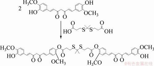
ͼ1�������ض�����ϳ�·��ͼ
Fig. 1��Synthesis route of curcumin dimer
ͨ���������(FTIR)�ͺ˴Ź������� (1HNMR)���������ض����壬�ֱ���ͼ2��ͼ3��ʾ��FTIR����(ͼ2)������1 627 cm-1�����ķ����ʻ���C=O�����йأ�1 592��1 507��1 429 cm-1�����ķ��DZ�����C=C����������ġ���CUR2-TK�Ĺ����У���1 730 cm-1���������µ����շ壬���������γ������C=O���������¡������ض�����CUR2-TK��1HNMR��(ͼ3)�������ں˴Ź��������У��ڻ�ѧλ��6~8֮��������д���CUR������ŵĶ�����������ӹ���壬��ѧλ��2.5���ķ�Ϊ뮴�DMSO�ܼ��壬��ѧλ��3.3���ķ�Ϊˮ�壬����Ϊ������м����ӻ��ŵ�����ͪTK��ͷ������������ڻ�ѧλ��1.62��2.92����������ɹ��ϳ���Ŀ����ͨ��ɫ���������������ض�����CUR2-TK�IJ���Ϊ76.37%��

ͼ2��CUR2-TK�ĺ������ͼ
Fig. 2��FTIR spectrum of CUR2-TK
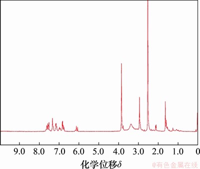
ͼ3��CUR2-TK�ĺ˴Ź�������ͼ
Fig. 3��1H NMR spectrum of CUR2-TK
2.2�����������Ʊ�����������ò����
������Ϊ��ˮ��ҩ���ˣ��Ʊ�(CUR2-TK)PEG-PLGA����������ˮ����(O/W)����Һ�ܼ��ӷ��������������Ʊ������У�ҩ�ェ���ض�����������PEG-PLGA�������ȡ�����DMSO����ȼ�������������������ǿ����ʱ�䡢ˮ��ɢ�����ȶ���PVA�����Լ�����ӷ��л��ܼ���ʱ������ض���Ӱ�������Ʊ������������ʣ�Ϊ����ͨ��EPRЧӦ��Ч��ʵ��������λ��ҩ����ۣ�����ǿ����������������������Ҫ�ϸ����������������������Ϊ50~100 nm���������ܴﵽ��ѵ�������ȡЧ�ʡ�ͨ����ʵ��������̽����ȷ���õ�����������������������£�����ۺ���PEG-PLGA�ڶ��ȼ����ܼ��е���������Ϊ2.2%��ˮ��ɢ����PVA����������Ϊ1%��������ˮ����������Ϊ1.3:10���г�������3 min���ټ�����������Ϊ0.1%��PVAˮ��Һ����600 r/min�Ľ����ٶȻӷ��л��ܼ�4 h�����Եõ�����С��100 nm����������
ͨ����羵�۲��Ʊ���(CUR2-TK)PEG-PLGA��������������ˮ��Һ��TEM��Ƭ��ͼ4��ʾ����ͼ4���Է��֣�������Ϊ���νṹ����ΪԲ�������ȽϾ�һ������������ȶ��ġ������������ε���������ͼ4(a)��ʾΪ��m(CUR2-TK):m(PEG-PLGA)=1:2�������Ʊ�������������羵ͼ����ͼ4(a)���Է�����������ƽ������ԼΪ40 nm������m(CUR2-TK):m(PEG-PLGA)��1:2�����ӵ�3:1ʱ����������ƽ�����������ӵ�76 nm���ң������������ˮ��ҩ�ェ���ض���������ӣ��Ʊ���������������֮����
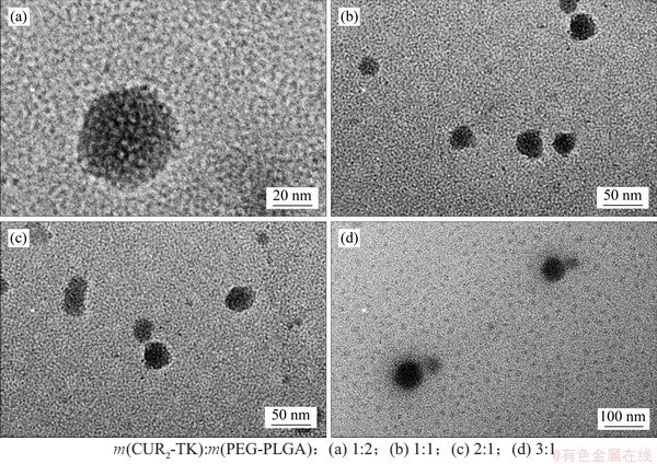
ͼ4����ҩ��������TEMͼ��
Fig. 4��TEM images of (CUR2-TK) PEG-PLGA NPs
�������������ȷ�����(Malvern Zeta-sizer Nano)���Ʊ�����������ˮ����������ɢϵ�����б�����DLS�������������ͼ5��ʾ����������羵�۲쵽��������������Ϊ�Ǻϡ���m(CUR2-TK):m(PEG-PLGA)�ֱ�Ϊ1:2��1:1��2:1��3:1������£�ƽ��ˮ�������ֱ�Ϊ(41.0��2.4)��(47.8��3.1)��(58.3��4.5)��(76.6��5.3) nm����������ҩ�ェ���ض����庬�������ӣ���������������֮����ͼ6��ʾΪ���ɢϵ��PDI��ҩ�ェ���ض����庬�����Ӷ�����ͬͶ�ϱ��µĶ��ɢϵ���ֱ�Ϊ0.168��0.009��0.215��0.013��0.323��0.021��0.409��0.025������������ҩ�����ӣ�������һ�Ա�
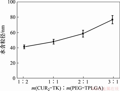
ͼ5����ͬҩ����μ�Ͷ�ϱ���������������
Fig. 5��Size with different mass ratios of CUR2-TK and PEG-PLGA
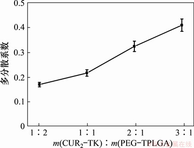
ͼ6����ͬҩ����μ�Ͷ�ϱ����������Ķ��ɢϵ��(PDI)�仯
Fig. 6��PDI with different mass ratios of CUR2-TK and PEG-PLGA
2.3������������ҩ���������
��ˮ��ҩ�ﵥ�帺���ھۺ������ҩ��ͨ��Ϊ10 %���ң�������Ҳ����ˡ�����ҩ�ﵥ��ϳɶ��������ҩ���ɴ�����ӵ�50%���ϡ���ˣ�ͨ��ҩ�ェ���ض�����CUR2-TK��ۺ���PEG-PLGA�Բ�ͬ���������Ʊ�������������ҩ�����������m(CUR2-TK):m(PEG-PLGA)�ı仯���1��ʾ��
��1����ͬCUR2-TK��PEG-PLGAͶ�ϱ�����������ҩ���Ͱ�����
Table 1��Drug loding and encapsulation efficiency with different mass ratios of CUR2-TK and PEG-PLGA
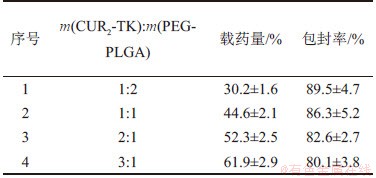
Ӱ������������ҩ��������ʵ���Ҫ����Ϊҩ��CUR2-TK��ۺ���PEG-PLGA��Ͷ�ϱȡ�Ϊ�����ҩ�����ҩ�������ٸ��μ���ʹ�ü��䶾�ԣ���Ҫ�ڼӴ�ҩ��ռ�ȵ�ͬʱ��֤һ���İ����ʡ���ͬCUR2-TK��PEG-PLGAͶ�ϱ��µ���������ҩ���Ͱ����ʼ���1���ӱ�1��֪������CUR2-TK���������ӣ�����������ҩ������ߣ�����������֮���͡�����ܹ�����CUR2-TK����������ʹO/W��ϵ��CUR2-TK��Ũ���ݶ���������Ũ���ݶȵ����ã������ҩ��CUR2-TK��ɢ��ˮ���δ�����������������ҩ����ʧ������ζ����m(CUR2-TK):m(PEG-PLGA)���ӣ�ҩ����O/W��ϵ�е�Ũ��������������ʼ�С�������ʾ����ҩ����ۺ���Ͷ��������Ϊ3:1ʱ����ҩЧ�ʸߴ�(61.9��2.9)%��Զ�����潪���ص��幹����10%���ҵ���ҩ��[5, 19-20]��������Ϊ(80.1��3.8)%���仯�����ԣ��Ҹ�Ͷ�ϱ��µõ��������������Ͼ�һ��ƽ������Ϊ76 nm��������ʵ���������Ʊ��Ľ����ض��������������бȽϺõ�Ӧ��DZ����
2.4��������������ҩ���ͷ�����
(CUR2-TK)PEG-PLGA��������������ҩ�о���pHΪ7.3ʱ�����Ỻ����Һ�н��У�����m(CUR2-TK):m(PEG-PLGA)=3:1�������ҩ�о���ͼ7(a)��ʾΪ�������ۻ�ҩ���ͷŶ�Qt��ʱ��t�����Թ�ϵ����ͼ7(a)��֪���ڿ�ʼ�Σ�����������Ĥ����ߵ�ҩ��Ũ�Ȳ����ҩ��Ѹ����ɢ����ҩ���ʴﵽ���壻����Ũ�Ȳ��С��ת��Ϊ���ͽΣ���100 h��ҩ���ۻ�ҩ���ͷŶ�Qt�ﵽ(84.87��3.15)%��
ͼ7(b)��ʾΪt1/2���ۻ�ҩ���ͷŶ�Qt�����ֱ�ߡ���ͼ7(b)��֪����Higuchi��ҩģ������Ժܺã���ҩģ����Ͻ�������������ڴ˻���Һ�е��ͷŷ���Higuchi���̣�Qt=14.45+6.95 t1/2(���ϵ��R=0.998)��
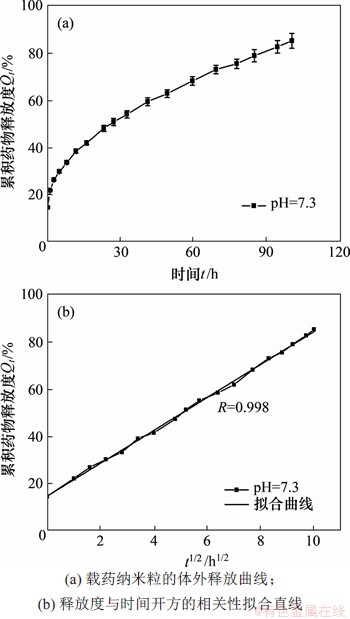
ͼ7����ҩ�������������ͷ����ߺ����ֱ��
Fig. 7��Release profiles of CUR2-TK from drug loaded nanoparticles and linear fitting chart
3 ����
1) �ϳ��˽����ض�����CUR2-TK�����Դ�Ϊҩ�ﵥԪ���Ծۺ���PEG-PLGA��Ϊҩ�����壬ͨ������Һ�ܼ��ӷ����Ʊ���(CUR2-TK)PEG-PLGA������������
2) ̽�����������Ʊ������в�ͬʵ�����������������ܵ�Ӱ�죬ȷ�����Ż����ʵ������������˽�������ҩ����ͨ��Ͷ�ϱȵ��ڣ���������ҩ���Ͱ����ʷֱ�ﵽ(61.9%��2.9)%��(80.1%��3.8)%����������ò�����Ͼ�һ��ƽ������Ϊ76 nm���ң���ҩ�ﻺ��ʱ���4 d���ϡ�
3) (CUR2-TK)PEG-PLGA��������Ч����˽����ص���ҩ���������������ʹ�ü����ܵĶ��ԣ��������ɿ�����50~100 nm֮�䣬����ǿ������λ���۵�ͬʱ�н�ǿ��������������������Ϊһ�ֿ�������ҩ�ﴫ��ϵͳ��
�ο����ף�
[1] NGAI S C. Curcumin sensitizes cancers towards TRAIL-induced apoptosis via extrinsic and intrinsic apoptotic pathways[J]. Current Drug Targets, 2020, 21: DOI:10.2174/1389450121666200302124426.
[2] HU Yuzhu, HE Yihong, JI Jianrui, et al. Tumor targeted curcumin delivery by folate-modified MPEG-PCL self-assembly micelles for colorectal cancer therapy[J]. International Journal of Nanomedicine, 2020, 15: 1239-1252.
[3] MUKHOPADHYAY R, SEN R, PAUL B, et al. Gemcitabine Co-encapsulated with curcumin in folate decorated PLGA nanoparticles��a novel approach to treat breast adenocarcinoma[J]. Pharmaceutical Research, 2020, 37(3): 56.
[4] YANG Jingzhe, WANG Chengli, ZHANG Zhijie, et al. Curcumin inhibits the survival and metastasis of prostate cancer cells via the Notch-1 signaling pathway[J]. Apmis, 2017, 125(2): 134-140.
[5] MAITI C, PARIDA S, KAYAL S, et al. Redox-responsive core-cross-linked block copolymer micelles for overcoming multidrug resistance in cancer cells[J]. ACS Applied Materials & Interfaces, 2018, 10(6): 5318-5330.
[6] CAI Kaimin, HE Xi, SONG Ziyuan, et al. Dimeric drug polymeric nanoparticles with exceptionally high drug loading and quantitative loading efficiency[J]. Journal of the American Chemical Society, 2015, 137(10): 3458-3461.
[7] PEI Qing, HU Xiuli, LIU Shi, et al. Paclitaxel dimers assembling nanomedicines for treatment of cervix carcinoma[J]. Journal of Controlled Release, 2017, 254: 23-33.
[8] PEI Qing, HU Xiuli, WANG Lei, et al. Cyclodextrin/paclitaxel dimer assembling vesicles: reversible morphology transition and cargo delivery[J]. ACS Applied Materials & Interfaces, 2017, 9(32): 26740-26748.
[9] FANG Shuo, HOU Yongpeng, LING Longbing, et al. Dimeric camptothecin derived phospholipid assembled liposomes with high drug loading for cancer therapy[J]. Colloids and Surfaces B: Biointerfaces, 2018, 166: 235-244.
[10] LI S D, HUANG L. Nanoparticles evading the reticuloendothelial system: role of the supported bilayer[J]. Biochimica et Biophysica Acta(BBA)-Biomembranes, 2009, 1788(10): 2259-2266.
[11] MATSUMOTO Y, NICHOLS J W, TOH K, et al. Vascular bursts enhance permeability of tumour blood vessels and improve nanoparticle delivery[J]. Nature Nanotechnology, 2016, 11(6): 533-538.
[12] ZHOU Chen, LONG M, QIN Yanping, et al. Luminescent gold nanoparticles with efficient renal clearance[J]. Angewandte Chemie International Edition, 2011, 50(14): 3168-3172.
[13] CHOI H S, LIU Wenhao, MISRA P, et al. Renal clearance of quantum dots[J]. Nature Biotechnology, 2007, 25(10): 1165-1170.
[14] TANG Li, YANG Xujuan, YIN Qian, et al. Investigating the optimal size of anticancer nanomedicine[J]. Proceedings of the National Academy of Sciences of the United States of America, 2014, 111(43): 15344-15349.
[15] MAEDA H, WU J, SAWA T, et al. Tumor vascular permeability and the EPR effect in macromolecular therapeutics: a review[J]. Journal of Controlled Release, 2000, 65(1/2): 271-284.
[16] YUAN Youyong, LIU Jie, LIU Bin. Conjugated-polyelectrolyte-based polyprodrug: targeted and image-guided photodynamic and chemotherapy with on-demand drug release upon irradiation with a single light source[J]. Angewandte Chemie International Edition, 2014, 53(28): 7163-7168.
[17] PEI Qing, HU Xiuli, ZHENG Xiaohua, et al. Light-activatable red blood cell membrane-camouflaged dimeric prodrug nanoparticles for synergistic photodynamic/chemotherapy[J]. ACS Nano, 2018, 12(2): 1630-1641.
[18] PHAECHAMUD T, TUNTARAWONGSA S. Transformation of eutectic emulsion to nanosuspension fabricating with solvent evaporation and ultrasonication technique[J]. International Journal of Nanomedicine, 2016, 11: 2855-2865.
[19] LIU Lisha, BI Yunke, ZHOU Muru, et al. Biomimetic human serum albumin nanoparticle for efficiently targeting therapy to metastatic breast cancers[J]. ACS Applied Materials & Interfaces, 2017, 9(8): 7424-7435.
[20] YAO Qing, DAI Zhi, CHOI J H, et al. Building stable MMP2-responsive multifunctional polymeric micelles by an all-in-one polyme-lipid conjugate for tumor-targeted intracellular drug delivery[J]. ACS Applied Materials & Interfaces, 2017, 9(38): 32520-32533.
(�༭ ����ƽ)
�ո����ڣ� 2020 -04 -01; �����ڣ� 2020 -06 -05
������Ŀ(Foundation item)������ʡ��Ȼ��ѧ����������Ŀ(2020JJ4680)������ʡ�о�������̽��������Ŀ(CX20190184)����������Ӣ����Ŀ(2018RS3005)�����ϴ�ѧ������Ӣ�ƻ���Ŀ(CX20190242) (Project(2020JJ4680) supported by the Natural Science Foundation of Hunan Province; Project(CX20190184) supported by the Graduate Independent Exploration and Innovation of Hunan Province; Project(2018RS3005)supported by Huxiang Youth Talent; Project(CX20190242)supported by the Sublimation Education of Central South University)
ͨ�����ߣ����ɣ���ʿ�������ڣ�����ҩ�ﻯѧ��ҩ���Ƽ��о���E mail: liuyf@csu.edu.cn
ժҪ��Ϊ����߿�����ҩ�ェ������ҩЧ�ʣ��Խ�����Ϊ��Ԫ�ϳ����ͽ����ض�����(CUR2-TK)�����Ծ��Ҷ���-�������ǻ����Ṳ����(PEG-PLGA)Ϊ���壬ͨ������Һ�ܼ��ӷ������Ʊ������ض����建�����������о���ͬҩ��CUR2-TK��ۺ���PEG-PLGA��������(m(CUR2-TK):m(PEG-PLGA))�ȶ����������ܵ�Ӱ�졣�о����������ͨ�������ض����幹������ҩ�������߱����ߵ���ҩЧ�ʣ���m(CUR2-TK):m(PEG-PLGA)Ϊ3:1ʱ����ҩ���Ͱ����ʷֱ�ﵽ(61.9��2.9)%��(80.1��3.8)%������������ò������һ�������ɿ���50~100 nm֮�䣬��ҩʱ���4 d���ϡ�
