
Bioactivity of bioresorbable composite based on bioactive glass and poly-L-lactide
ZHOU Zhi-hua(周智华)1, 2, RUAN Jian-ming(阮建明)1, ZOU Jian-peng(邹俭鹏)1,
ZHOU Zhong-cheng(周忠诚)1, SHEN Xiong-jun(申雄军)1
1.State Key Laboratory of Powder Metallurgy, Central South University, Changsha 410083, China;
2.College of Chemistry and Chemical Engineering, Hunan University of Science and Technology,
Xiangtan 411201, China
Received 7 June 2006; accepted 28 November 2006
Abstract: Bioactive and bioresorbable composite was fabricated by a solvent evaporation technique using poly-L-lactide(PLLA) and bioactive glass (average particle size: 6.8 μm). Bioactive glass granules are homogeneously distributed in the composite with microcrack structure. The formation of hydroxyapatite(HA) on the composite in simulated body fluid(SBF) was analyzed by scanning electron microscopy(SEM), energy dispersive spectroscopy(EDS), X-ray diffraction(XRD), and Raman spectra. Rod-like HA crystals deposit on the surface of PLLA/bioactive glass composite after soaking for 3 d. Both rod-like crystals and HA layer form on the surface for 14 d in SBF. The high bioactivity of PLLA/bioactive glass composite indicates the potential of materials for integration with bone.
Key words: bioactivity; composite; bioactive glass; poly-L-lactide
1 Introduction
Bioabsorbable polymeric implants, such as poly-L-lactide(PLLA), have been extensively used in the treatment of traumas of the skeletal system[1]. Synthetic bioresorbable polymers, in particular PLLA, polyglycolic and their copolymers, are successfully used as bio- materials. They can retain the tissue supporting property for a given length of time, and then gradually biodegrade [2]. A major drawback of these materials, however, is the release of acidic degradation products that may lead to inflammatory responses[3]. Another limitation is their lack of bioactivity, which means, for the case of bone tissue engineering, that they do not allow bone apposition or bonding on the polymer surface[4].
Some bioceramics, such as bioactive glass and calcium phosphates, are osteoconductive and thus can effectively accelerate bone tissue healing[5-6]. Biodegradability and bioactivity can be combined in the form of composites to obtain optimized materials exhibiting tailored mechanical propertied and controllable resorption rates in the body[7]. In the past few years, increasing attention has been paid to composites of bioabsorbable or biostable polymers and bioceramics. Various approaches to the development of such composites are investigated worldwide[8-14]. Usually bioceramics are combined with the bulk or porous polymer matrix either as fillers or as coatings.
In the present work, a solvent evaporation technique was used to prepare PLLA/bioactive glass composites. The effects of bioactive glass on the microstructure and bioactivity of the composites were studied. The potential bone integration ability of the PLLA/bioactive glass composites was demonstrated by a model of simulated body fluid(SBF) in vitro.
2 Experimental
2.1 Materials and processing
L-lactide, prepared in our laboratory, was used as a monomer for PLLA synthesis. Stannous octoate was used as an initiator without additional purification. Ampoules for polymerization were dried overnight in an oven at 400 ℃. Then, they were loaded with freshly recrystallized L-lactide and Sn-octoate toluene solution with molar ratio of monomer to initiator of 1 000-12 000, sealed under high vacuum, and left in the oven at 140 ℃ so that the material could polymerize. The polymerization time varied from 12 to 60 h. After the reaction was finished, the polymer was dissolved in chloroform, precipitated in methanol and dried in vacuum to constant mass. PLLA had a viscosity-average relative molecular mass about 4.0×10⁵. Bioactive glass with an average particle size of 6.8 μm and a composition of 35CaO, 60SiO2, 5P2O5 (mole fraction, %) was also prepared by sol-gel method in our laboratory according to Ref.[15]. The XRD study of the bioactive glass confirms an amorphous material (Fig.1).

Fig.1 XRD pattern of bioactive glass
PLLA/bioactive glass composite was prepared in our lab. Briefly, the polymer was dissolved in CH2Cl2 to produce a polymer solution (0.05 g/mL). The mixture was stirred overnight to obtain a homogeneous polymer solution. Bioactive glass powder (10%, mass fraction) was added into the polymer solution. The mixture was transferred into a flask and sonicated for 60 min in order to improve the dispersion of the bioactive glass particles into the polymer solution. After homogenization, the mixture was cast onto a flat glass plate to obtain flake then heated in a vacuum drying oven at 40 ℃.
2.2 In vitro studies and characterization
In vitro test was carried out by soaking disks of the glass in a so-called SBF. The SBF was prepared by dissolving reagent chemical of NaCl, NaHCO3, KCl, K2HPO4・3H2O, MgCl2・6H2O, CaCl2, and Na2SO4 into deionized water[16]. The fluid was buffered at physiological pH of 7.32 at 37 ℃ with tris (hydro- xymethyl) aminomethane and hydrochloric acid. The inorganic composition of SBF emulated that of human blood plasma. Two pieces of each specimen were used to investigate the reaction of sol-gel glass to SBF. Each specimen was immersed in shaken SBF (A/V=0.1 cm2/mL, 60 r/min). The samples were immersed in the SBF fluid for 3, 7, 10 and 14 d at a constant temperature of 37 ℃.
Bioactive glass particle size and size distribution of the resulting powder were analyzed by Laser Scattering Particle Size Distribution Analyzer (Microplus). After soaking, specimens were taken from the fluid, rinsed with deionized water, and dried at 37 ℃ for scanning electron microcopy connected with energy dispersive spectrocopy (SEM-EDS), X-ray diffraction(XRD) and Raman spectra. The SEM-EDS study was made on a JSM-6360LV microscope. For the SEM study, the pieces were coated with a film of gold. XRD patterns were obtained in a Philips X’Pert MPD diffractometer using Cu Kα radiation in θ-2θ scans and grazing incidence 2θ scans. The Raman spectrum was obtained using JOBIN YVON HORIBA Raman spectroscopy. Ionic concentrations in SBF were measured by inductively coupled plasma-atomic emissive spectrum (ICP-AES).
3 Results and discussion
3.1 Morphology of polymer/bioactive glass composite
The surface morphology of composite is shown in Fig.2. Bioactive glass is prepared with the average size of 6.8 μm, but there exists the agglomeration of bioactive glass particles. The SEM micrograph provides evidence of the existence of a small percentage of large glass particles (>10 μm). The bioactive glass particles, small or large, are angular in shape. Bioactive glass particles are distributed evenly in the composite nevertheless the polymer covers the bioactive glass particles on the surface (Fig.2(a)). As shown in the magnification micrograph (Fig.2(b)), the polymer has a microcrack structure due to the shrinkage of the polymer caused by the evaporation of CH2Cl2. The microcrack structure will weaken the structure of the composite; however, it will be of benefit to bioactivity of composites because of larger surface area.

Fig.2 SEM micrographs of PLLA/bioactive glass before soaking in SBF
3.2 Changes in SBF
The variation of pH value relative to soaking times in SBF of composite is shown in Fig.3.

Fig.3 Variation of pH value in SBF of PLLA/bioactive glass
The increasing pH value during the first hours about 50 h due to partial dissolution at the surface level indicates the high reactivity of these materials. These facts agree with the formation mechanisms of the apatite on bioactive glasses and glass ceramics; i.e., in early stages an interchange takes place between Ca2+ and H3O+ from the solution. Such interchanges provoke an increase of the pH that favors the formation of apatite nuclei on the silanol groups on the glass surface. In this interval a fluctuation in the pH of PLLA/bioactive glass is also detected, which may be explained if we consider the result of two opposite processes: the release of Ca2+ from the glass and the consumption of Ca2+ due to formation of the apatite-like layer. Therefore, when the release rate of Ca2+ is higher than the consumption rate, the pH will increase, or else, the pH value will decrease. Compared with incubation of sol-gel bioactive glass, pH value of composites increases slowly during immersion and is lower than 8.0[17]. In the case of polymer/bioactive glass composite, the bioactive glass should act as a protective hydrolysis barrier affecting the rate of degradation of the polymer substrate, as found in recent investigations[18-19]. The rapid exchange of protons in water for alkali in the glass provides a pH value buffering effect on the polymer surface, therefore acceleration of degradation will not occur due to small pH value changes during bioactive glass dissolution.
During the immersion, the concentration of P in solution decreases, while the concentrations of Ca and Si increase. As can be seen in Fig.4, during the first 24 h Ca and Si concentrations increase from 95 to 150 μg/g and from 0 μg/g to 5.2 μg/g, respectively, whereas P concentration decreases from 30 to 18.2 μg/g. The concentration of these ions in SBF changes dramatically during the first hours of immersion. Then, the variation slows down gradually. The continuous decrease of P concentration in solution during the first hours indicates that the amount of P released from glass can’t compensate its consumption caused by apatite deposition. After 7 d soaking, the ionic concentration of P and Si remained without variation with respect to the values obtained after 3 d whereas Ca ionic concentration increases up to 140.7 μg/g. The results for the release of silica do not correspond with previous studies of polysulfone/bioactive glass compositions[20].

Fig.4 Variation of Ca, P, and Si ionic concentration with immersion in SBF for PLLA/bioactive glass
3.3 Formation of apatite-like layer
The deposition on the surface of PLLA/bioactive glass composite in SBF was analyzed qualitatively using SEM-EDS, Raman spectrum and XRD.
The XRD patterns of the surface of composite before and after immersion in SBF are shown in Fig.5. The XRD study of the composite confirms a low crystalline polymeric composite material, with two peaks at 16.63? and 18.86? corresponding to the (200) and (203) reflection of PLLA respectively, in the samples before soaking. A slight sharpening of the broad peak is observed in the XRD pattern of the glass after soaking for 3 d. The trace of the surface immersion shows diffraction peak at 32.6? corresponding to the (211) reflection of calcium phosphate hydroxide (HA). The apatite diffraction peak at 39.8?, corresponding to (222) reflections of HA, also becomes very sharp in the XRD pattern of the glass after 7 d immersion. Other less intense peaks at 46.8? corresponding to (310) reflections of HA respectively are also identified after 14 d immersion.

Fig.5 XRD patterns of PLLA/bioactive glass before and after soaking in SBF
Fig.6 presents the gradual development of HA on the surface as a result of increasing days of immersion in SBF, i.e. after 3, 7, 10 and 14 d, as confirmed by XRD analyses.
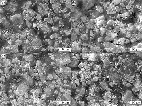
Fig.6 SEM micrographs of composites after 3, 7, 10 and 14 d soaking in SBF
Fig.6 shows the morphology of the composite surfaces changes after 3 d immersion. Small rod-like hydroxyapatite crystals have deposited on the surface, which is different from the morphology of porous
polymer/bioactive glass[21-22]. The rod-like HA crystals, with priority growth, increase in quantity and lengthen with increasing the immersion time in SBF. As shown in Fig.7, the rod-like crystal grows from bioactive glass and then dendrite forms. The EDS of the crystal shows that it is composed of calcium, phosphor and little silicon after 7 d soaking in SBF (Fig.7(A)), which confirms the formation of HA. After 7 d soaking in SBF, the EDS analysis of bioactive glass surface can be detected a clear increase in calcium and phosphate compared with the silicon concentrations. Figs.7(C) and (D) indicate both rod-like crystals and HA layer are formed on the surface after 14 d immersion because the silicon concentration is little and the calculated Ca/P ratio is about 1.8, which is comparable to the Ca/P ratio of the human bone, as it ranges between 1.6 and 1.7 depending on the age of the person.

Fig.7 High-magnification SEM images after 7, 14 d immersion and EDS of PLLA/bioactive glass after immersion
The formation of HA on the surface of the composite with increasing days of immersion in SBF is confirmed by the Raman spectroscopy (Fig.8). For comparison, the spectrum for a sample under the as-fabricated condition is also shown. The formation of the HA on the surface of PLLA/bioactive glass is indicated by the strong peak at 960 cm-1, which corresponds to the symmetric stretching vibration of P―O in  tetrahedral belonging to hydroxyapatite crystals. The peak at 960 cm-1 increases with increasing immersion time in SBF, which indicates that the amount of HA formed on the sample increases with time. The 875 cm-1 peak in the Raman spectrum, corresponding to C―COO stretching in PLLA, decreases with increasing immersion time in SBF. Therefore the ratio between the heights of the peaks at different immersion time may be used to quantify the relative amount of HA formed during immersion in SBF.

Fig.8 Raman spectra of PLLA/bioactive glass before and after immersion
It is well known that bioactive glass has a higher index of bioactivity than that of HA, which makes it a more suitable material for applications in bone reconstruction[23]. It has been shown, for example, that there is much more bone formed in one week in the presence of bioactive glass than that is formed when HA or other calcium phosphate ceramic particulates are placed in the same type of defect[24]. Moreover bioactive glass, being a class A bioactive material (as opposed to HA, which is class B), has shown strong bond also to soft tissues[25]. The application potential of the bioactive glass-containing composites fabricated here, therefore, could include both hard and soft tissue regeneration and repair.
The rapid release of calcium ions into the solution, as well as the pH increases during the first hours of the essay as a consequence of partial dissolution at the surface level, giving an idea of the high reactivity of these materials. These facts agree with the formation mechanisms of the apatite-type layer on bioactive glass [26]. A complex process for the formation of apatite layer, involving five reaction stages, has been proposed in early stages and an interchange takes place between alkaline- earth ions from the substrate with H3O+ from the solution. Such interchanges provoke an increase of the pH that favors the formation of apatite nuclei on the silanol groups on the glass surface. Loss of soluble silica in form of Si(OH)4 to the solution results from breaking of Si―O―Si bonds and forms Si―OH at the glass solution interface. Then, there are condensation and repolymerization of an SiO2-rich layer on the surface depleted in alkaline earth cations. Migration of Ca2+ and  groups to the surface through the SiO2-rich layer takes place and forms a CaO-P2O5-rich film on top of the SiO2-rich layer, followed by growth of the amorphous CaO-P2O5-rich film by incorporation of soluble calcium and phosphates from solution. Finally, crystallization of the amorphous CaO-P2O5-rich film takes place by incorporation of OH- or
  anions from solution to form a mixed hydroxyl, carbonate layer.
4 Conclusions
1) Bioactive and bioresorbable PLLA/bioactive glass was developed based on PLLA and bioactive glass particles. Bioactive glass particles are distributed homogeneously in the composite, and the polymer has a microcrack structure due to the evaporation of CH2Cl2.
2) Rod-like HA crystals deposit on the surface of PLLA/bioactive glass composite after 3 d immersion. Both rod-like crystals and HA layer are formed on the surface after 14 d immersion.
3) The calculated Ca/P ratio of HA is about 1.8, which is comparable to the Ca/P ratio of the human bone. The composite containing bioactive glass is potential implant material for small bone fracture fixations.
References
[1] ROKKANEN P U, BOSTMAN O, HIRVENSALO E. Bioabsorbable fixation in orthopaedic surgery and traumatology [J]. Biomaterials, 2000, 21(24): 2607-2613.
[2] DE JONG W H, EELCO B J, ROBINSON J E. Tissue response to partially in vitro predegraded poly-L-lactide implants [J]. Biomaterials, 2005, 26(14): 1781-1791.
[3] AGRAWAL C M, RAY R B. Biodegradable polymeric scaffolds for musculoskeletal tissue engineering [J]. Biomed Mater Res, 2001, 55(2): 141-150.
[4] WEIR N A, BUCHANANA F J, ORRA J F. Processing, annealing and sterilisation of poly-l-lactide [J]. Biomaterials, 2004, 25(18): 3939-3949.
[5] VALLET-REGI M, RAGEL CV, SALINAS A J. Glasses with medical applications [J]. Eur J Inorg Chem, 2003, 6(12): 1029-1042.
[6] VALLET-REGI M. Ceramics for medical applications [J]. Chem Soc Dalton Trans, 2001, 17(1): 97-108.
[7] ROETHER J A, BOCCACCINIB A R, HENCH L L. Development and in vitro characterisation of novel bioresorbable and bioactive composite materials based on polylactide foams and bioglass for tissue engineering applications [J]. Biomaterials 2002, 23(19): 3871-3878.
[8] NENAD I, DRAGAN U. Synthesis and application of hydroxyapatite/polylactide composite biomaterial [J]. Applied Surface Science, 2004, 238(1-4): 314-319.
[9] BOCCACCINI A R, ROETHER J A, HENCH L L. Composites approach to tissue engineering [J]. Ceram Eng Sci Proc, 2002, 23(5): 805-816.
[10] SULJOVRUJIC E, IGNJATOVIC N, USKOKOVIC D. Gamma irradiation processing of hydroxyapatite/poly-L-lactide composite biomaterial [J]. Radiation Physics and Chemistry, 2003, 67(3/4): 375-379.
[11] SOICHIRO I, MASANORI K, YOSIHISA K. Development of an artificial vertebral body using a novel biomaterial, hydroxyapatite/collagen composite [J]. Biomaterials, 2002, 23(19): 3919-3926.
[12] NIEMELA T, KELLOMAKI M, TORMALA P. In vitro degradation of osteoconductive poly-L/DL-lactide/b-TCP composites [J]. Key Eng Mater 2004, 54(3): 509-512.
[13] NIIRANEN H, PYHALTO T, ROKKANEN P. In vitro and in vivo behavior of self-reinforced bioabsorbable polymer and self-reinforced bioabsorbable polymer/bioactive glass composites [J]. Biomed Mater Res, 2004, 69A(6): 699-708.
[14] JUNG Y M, KIM S S, KIM Y H. A poly(lactic acid)/calcium metaphosphate composite for bone tissue engineering [J]. Biomaterials, 2005, 26(32): 6314-6322.
[15] XIA Wei, CHANG Jiang. Well-ordered mesoporous bioactive glasses (MBG): A promising bioactive drug delivery system [J]. Journal of Controlled Release, 2006, 110(3): 522-530.
[16] SARAVANAPAVAN P, HENCH L L. Low-temperature synthesis, structure, and bioactivity of gel-derived glasses in the binary CaO-SiO2 system [J]. Biomed Mater Res, 2001, 54(6): 608-618.
[17] SEPULVEDA P, JONES J R, HENCH L L. In vitro dissolution of melt-derived 45S5 and sol-gel derived 58S bioactive glasses [J]. Biomed Mater Res, 2002, 61(3): 301-311.
[18] STAMBOULIS A, HENCH L L. Bioresorbable polymers: Their potential as scaffolds for bioglasss composites [J]. Key Eng Mater, 2001, 192(7): 729-732.
[19] STAMBOULIS A, BOCCACCINI A R, HENCH L L. Novel biodegradable polymer/bioactive glass composites for tissue engineering applications [J]. Adv Eng Mater, 2002, 4(2): 105-109.
[20] MARCOLONGO M, DUCHEYNE P, LACOURSE W C. Surface reaction layer formation in vitro on a bioactive glass fiber/polymeric composite [J]. Biomed Mater Res, 1997, 37(3): 440-448.
[21] ROETHERA J A, BOCCACCINIB A R, HENCH L L. Development and in vitro characterization of novel bioresorbable and bioactive composite materials based on polylactide foams and bioglass for tissue engineering applications [J]. Biomaterials, 2002, 23(20): 3871-3878.
[22] MAQUET V, BOCCACCINIC A R, PRAVATAA L. Porous poly(a-hydroxyacid)/bioglass composite scaffolds for bone tissue engineering (I): Preparation and in vitro characterization [J]. Biomaterials, 2004, 25(21): 4185-4194.
[23] JONES J R, EHRENFRIED L M, HENCH L L. Optimising bioactive glass scaffolds for bone tissue engineering [J]. Biomaterials, 2006, 27(7): 964-973.
[24] HENCH L L, XYNOS I D, EDGAR A J, BUTTERY LD K, POLAK J M. Gene activating glasses [C]//Proceedings of the Nit Congr Glass, vol. 1. Sheffield, UK: Society of Glass Technology, 2001: 226-233.
[25] BORRAJO J P, GONZ??LEZ P, LISTE S. The role of the thickness and the substrate on the in vitro bioactivity of silica-based glass coatings [J]. Materials Science & Engineering C, 2005, 25(2): 187-193.
[26] VERN?? E, NUNZIO S DI, BOSETTI M. Surface characterization of silver-doped bioactive glass [J]. Biomaterials, 2005, 26(25): 5111-5119.
Foundation item: Project(50174059) supported by the National Natural Science Foundation of China
Corresponding author: RUAN Jian-ming; Tel: +86-731-8876644; E-mail: jianming@mail.csu.edu.cn
(Edited by LI Xiang-qun)

