���±�ţ�1004-0609(2011)08-1944-09
Co0.5Ni0.5Fe2O4������ά�ľ����˿��
�Ʊ����������������
�� ��1, 2��������1���ܹ���1��������1������ǭ2
(1. ���տƼ���ѧ ����ѧԺ���� 212003;
2. ���մ�ѧ ���Ͽ�ѧ�빤��ѧԺ���� 212013)
ժ Ҫ�������ܽ�-��������Ͼ����˿�����ɹ��Ʊ�ֱ��Ϊ150~400 nm��Co0.5Ni0.5Fe2O4������������ά��������TG-DTA��XRD��FTIR��FESEM��TEM��VSM����Ʒ���б��������������ǰ������ά��450 �決�պ�ɻ����γɴ��ྦྷ̬��Ŀ�����Co0.5Ni0.5Fe2O4������ά�����ű����¶ȵ����ߣ���άֱ����С�������ߴ���������ά����ֲڶȼ�ǿ��������ò�ɶ�ṹ������״�ṹת�䣻���Ƶõ�Co0.5Ni0.5Fe2O4������ά�ı��ʹŻ�ǿ����450 ��ʱ��35.8 A��m2/kg�������ӵ�1 000 ��ʱ��80.2 A��m2/kg����������������������С�����ƣ���550 �渽��ʱ�ﵽ���ֵ����ζ���䵥���ٽ�ߴ�ԼΪ30 nm���ҷ����ڵ���ߴ緶Χ�ڣ���������ƽ�������ߴ��0.7�η������ȣ��������������ģ����Ԥ��Ľ�������Ǻϡ����⣬����(77 K)�Ų�����ʾ����������ȣ���Ʒ�Ľ�������ʣ��Ż�ǿ�Ⱦ������ߣ������ʹŻ�ǿ�������½���
�ؼ��ʣ�Co-Ni�����壻������ά�������˿��������
��ͼ����ţ�TQ 343+.41; O 482.5���� ���ױ�־�룺A
Electrospinning fabrication, characterization and
magnetic properties of Co0.5Ni0.5Fe2O4 nanofibers
XIANG Jun1, 2, CHU Yan-qiu1, ZHOU Guang-zhen1, GUO Yin-tao1, SHEN Xiang-qian2
(1. School of Mathematics and Physics, Jiangsu University of Science and Technology, Zhenjiang 212003, China;
2. School of Materials Science and Engineering, Jiangsu University, Zhenjiang 212013, China)
Abstract: Co0.5Ni0.5Fe2O4 nanofibers with diameter of 150�C400 nm were fabricated by sol-gel method combined with electrospinning technology. The as-prepared fiber samples were characterized by means of TG-DTA, XRD, FTIR, FESEM, TEM and VSM techniques. The results show that the pure resultant products with spinel structure are basically formed after calcining the precursor fibers at 450 �� for 2 h. With the increase of the calcination temperature, the fiber diameters decrease and the mean size (D) of Co0.5Ni0.5Fe2O4 grains within the fibers gradually increase. As a result, the fiber surface morphology gradually transforms from a porous structure to a necklace-like structure. The saturation magnetizations (Ms) of the obtained Co0.5Ni0.5Fe2O4 nanofibers monotonously increase from 35.8 A��m2/kg at 450 �� to 80.2 A��m2/kg at 1 000 �� while the corresponding coercivities (Hc) initially increase and then decrease, and reaches the maximum value at about 550 ��, which indicates that the single-domain critical size of Co0.5Ni0.5Fe2O4 in the form of nanofibers is around 30 nm. Hc of the nanofibers varies as D0.71 in a D range below the single-domain critical size, which is in good agreement with the result predicted on the basis of the random anisotropy model. In addition, the magnetic properties measured at low temperature (77 K) show the substantial enhancements in both coercivity and remanent magnetization and a relatively obvious reduction in saturation magnetization compared to the corresponding room-temperature values.
Key words: Co-Ni ferrite; nanofibers; electrospinning; magnetic properties
��������ѧ�����Ŀ��ٷ�չ��������������ϵĺϳɡ����ܼ���Ӧ���ѳ�Ϊ��ǰ���ϡ���������ѧ��ѧ�Ƶ��о��ȵ�֮һ��������������ж��ص��ṹ�ʹ����ܣ���һ�����ܽ�Ϊ��Խ�Ĵ��Բ��ϣ��ѹ㷺Ӧ������Ϣ���ӡ��Ŵ洢�������塢�����䡢����������ҽѧ���������������Ÿ����Լ���Ų����յ�����[1-2]��CoFe2O4��һ�ּ⾧ʯ��Ӳ�Ų��ϣ����нϸߵı��ʹŻ�ǿ�ȡ��ž��������Ժͽ������Լ����õĻ�ѧ�ȶ��ԡ����������ԡ��ŵ��ԺʹŹ����ܵ��ŵ�, �ڴ�����ҩ�����塢���ܶ���Ϣ�洢�����Ծ�̬�������������沨�������͵���Ĵ�о���ϵ�����������Ҫ��Ӧ�ü�ֵ[3-6]��Ȼ��������CoFe2O4���нϴ�����Ĵž��������Գ���K1(3.8��105 J/cm3)�����������ýϸߵ���ʼ�ŵ��ʣ�����һ���̶����ƴ�����ϵ�Ӧ�á�Ϊ�����㲻ͬӦ�õ���Ҫ���Ե�Ԫ��������в��Ӹ��Լ�ͨ����������γɶ�Ԫ�������Ե�����硢�������ѳ�Ϊ��ǰ����������о���һ����Ҫ������NiFe2O4��һ�־��еͽ������ͱ��ʹŻ�ǿ���Լ����ø�Ƶ������Ե����Ų��ϣ���ž��������Գ���K1��С��Ϊ��ֵ(-6.7��103 J/cm3)����ˣ���Ni2+����ȡ��Co2+��������Ч�ض�CoFe2O4�Ĵž��������Գ���K1����������ϵ���ˡ�����糣���š����ŵ��ʦ̡�������Hc�ͱ��ʹŻ�ǿ��Ms�ȵ硢�Ų������е��أ��Ӷ����Ի�þ�������������ܵļ⾧ʯ������(Co-Ni)���������塣Ŀǰ�������Ѳ����ܽ�-������������������ˮ�ȷ������������ܽ�����ȼ�շ��������Ƚⷨ����ת��Ʒ������յ�ȼ�պϳɵȼ����Ʊ�������Co-Ni���������[7-13]�ͱ�Ĥ[14-15]����������δ���й�Co-Ni������һά��һά���ṹ���Ϸ�����о�������
��ʮ������һά��һά���Խṹ�����������ߡ����ܡ����״���������ά�ȱ��ܹ�ע���������Ǿ��нϸߵij����ȡ��ȱ��������״�������Ե����������ֳ���ͬ����Ӧ�����ͻ��ĵ������ͻ�ѧ���ܣ�ʹ�����ڳ����ܶ����ݴ洢���Ŵ��������ɵ���������������������������ҽѧ�Լ���Ų����յ�����չ�ֳ����˵�Ӧ��ǰ��[16-19]�������˿����ʼ��20����30��������ֻ�������Ʊ�һЩ�ۺ�����ά��ֱ�����ʮ�������ü����ű�Ӧ��������һά���ṹ���ϵ��Ʊ���[20-22]�������˿�������ѱ���Ϊ���Ʊ�����������ά���ʵ�õķ���֮һ����������øü����ɹ��غϳ���һЩ�⾧ʯ��������������ά�����״�[17-18, 23-26]�����������Ծ���ϩ������ͪ(PVP)Ϊ��ϼ�������Ӧ�Ľ��������η�Ӧ�Ƶ�ǰ������Һ���þ����˿���Ʊ�Co0.5Ni0.5Fe2O4������ά�������ö����Ƚ������ֶζԺϳ���Ʒ�Ľṹ������ò�ʹ����ܽ��б�����
1 ʵ��
1.1 ��ά�Ʊ�
�����ܽ�-��������Ͼ����˿�����Ʊ��⾧ʯ��Co0.5Ni0.5Fe2O4������ά�����Ʊ�������Ҫ��ǰ������Һ���ơ����˿���ȴ���3�����ֹ��ɡ��ȳ�ȡ0.808 5 g����ϩ������ͪ(PVP����������Aldrich��ƽ����Է�������Ϊ1 300 000) ����9.318 3 g��ˮ�Ҵ���2 gȥ����ˮ����ɵĻ���ܼ��У����������´�������Լ1 hʹPVP��ȫ�ܽ⡣Ȼ���ٰ�������Ԫ��n(Co):n(Ni):n(Fe)=1:1:4��ȡ0.181 9 g�����ܡ�0.155 3 g��������1.01 g������(��Ϊ����������ҩ���Ż�ѧ�Լ�����˾����)���뵽����PVP��Һ�У���������������Լ10~15 h���Ƶóɷ־��ȵ�ǰ������Һ�����н����ε�Ũ��Ϊ10%(��������)��PVPŨ��Ϊ6%(��������)��
������ǰ������Һ������в������ͷ (�⾶0.8 mm)������ע�����У�����װ����ע���(TS-1B/ W0109-1B�������������������˾)�ϣ��������ͷ���ѹ��Դ(RR30-5P/DDPM/220, Gamma)������������������Ϊ���������ѹ��Դ�ĸ�������(�����ӵ�)������Һ��������Ϊ0.5 mL/h�����ܾ���Ϊ15 cm����ѹΪ15 kV�������½��о����˿������������ռ����ij��������е�ǰ������ά��������ڳ̿ص�¯�У��ֱ���450��550��600��650��700��800��1 000 ���±���2 h���Ƶ�Ŀ�����Co0.5Ni0.5Fe2O4������ά�����������ʾ�Ϊ3 ��/min��
1.2 ������
����DTG-60H�Ͳ���-����(TG-DTA)ͬ���������о�ǰ������ά���ȷֽ���̣��¶ȷ�ΧΪ������900 �棬������Χ����������10 ��/min��ʹ��D/max 2500 PC��X����������(XRD)����ǰ������ά����ת�����̼�Ŀ��������ά�ľ���ṹ��X����ԴΪCu��(��=0.154 06 nm)��ɨ���ٶ�Ϊ6 (��)/min��ɨ�跶ΧΪ15��~70��(2��)������JSM-7001F�ͳ�����ɨ��羵(FE-SEM)��JEM-2100����羵(TEM)�۲����Ʊ���ǰ���弰����������ά����ò���۽ṹ��ʹ��Nicolet 380����Ҷ�任�������(FTIR)�Dz�����Ʒ�ĺ�����ף�����KBrѹƬ��������Χ400~4 000 cm-1������HH-15������Ʒ��ǿ��(VSM)������ά��Ʒ�����º͵���(Һ����77 K)�µĴ����ܡ�
2 ���������
2.1 TG-DTA����
ͼ1��ʾΪCo0.5Ni0.5Fe2O4/PVPǰ������ά��TG-DTA���ߡ���ͼ1�п��Կ�����DTA������������140 ��֮����һ��С�����ȷ壬��Ӧ��TG���߳���Լ10.5%��������ʧ����������ǰ������ά�в����ܼ��Լ���������������ˮ�Ļӷ���ɵġ���140~400 �������ڣ�DTA�����ϳ������������ȷ壬��Ӧ��TG������������������ʧ̨�ף�������ʧ����Ϊ70.5%���ڷ���270 �洦���۲쵽�Ŀ���ǿ�ķ��ȷ���Ҫ��ǰ������ά�н����ηֽ⡢PVP̼���ֽ��Լ�̼������ͬ�������£�����������Ӧ���̽�֯��һ����ʹ������Ӧ���̴�����������ʧ������һ�𣬴Ӷ�������һ���ϴ��������ʧ̨��[27]����362 �洦���ֵ�С�ķ��ȷ��������ά�в�������κ��л���Ľ�һ�������ֽ���ɵģ�400 ���Ժ�TG��DTA���߶�����ƽ�ȣ�˵����ʱǰ������ά���ȷֽ������������ȫ��ʣ���Ϊ�����������Ԥʾ�ż⾧ʯCoNi����������ܿ�ʼ�γɡ�
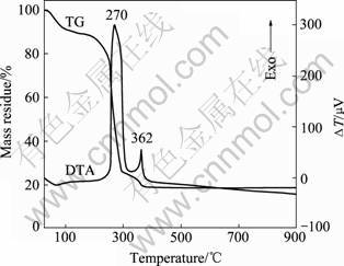
ͼ1 ǰ������ά��TG-DTA����
Fig.1 TG-DTA curves of precursor fibers
2.2 XRD����
ͼ2��ʾΪǰ������ά�����ڲ�ͬ�¶��±��� 2 h���ò����XRD�ס���ͼ2�ɼ�������ǰǰ������ά�����νṹ����2�� = 22�㸽�����ֵ�һ�������������ΪPVP�İ뾧���塣���ű����¶ȵ����ߣ�ǰ������ά�еĽ����κ��л����ֽ⣬��ṹ�ɷǾ�̬����̬ת�䣻��450 �決�պ��Թ۲쵽���Ե�����壬����������嶼�ɰ������⾧ʯ�ṹ����ָ�껯��������������֣�˵���ѻ����γɾ�̬��Co0.5Ni0.5Fe2O4������ά������ʱ��������ǿ����Խ��������νϿ���˵�����¶��ºϳɲ���Ľᾧ�̶Ȳ��ߡ������ߴ绹�Ƚ�С�����ű����¶ȵĽ�һ�����ߣ����Կ�����������ǿ�������ӣ������С������Խ��������ζ����ά�ľ����̶��Լ����������Զ�����ߣ������ߴ�Ҳ�������ܹ��õ�������Ϊ��õľ�̬Ŀ������������(311)������������ݣ�����Scherrer��ʽ�����������ά��Ʒ��ƽ�������ߴ磺
 (1)
(1)
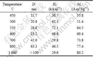
ʽ�У���Ϊ����X���ߵIJ���(1.540 6 ?)���ºͦȷֱ�Ϊ�������İ�߿��Ͳ�����ǣ����ý�����ڱ�1�С��ɱ�1���Կ��������ű����¶ȵ����ߣ����Ʊ���Co0.5Ni0.5Fe2O4������ά��ƽ�������ߴ���450 ��ʱ��11.7 nm��������1 000 ��ʱ��100 nm���ϡ�
2.3 FTIR����
ͼ3��ʾΪǰ������ά�����ڲ�ͬ�¶��±��ղ����FTIR�ס���ͼ3�п��Կ���������ǰ������ά�������г��ֵ����շ���Ҫ��PVP���ᾧˮ������ˮ�Լ������е�C��H��C��C��C��O��O��H��N��O�������������������շ�[28]����450 �決�պ�, �л���������״�������ʧ��ͬʱ�ڵͲ���595 cm-1�����������µ����շ壬��Ӧ�ڼ⾧ʯ������������λ��������(M��O)�����������շ�[29]��˵���ڸ��¶��£�ǰ������ά�ѻ�������������Ŀ�����Co0.5Ni0.5- Fe2O4������ά�ѻ����γɡ�����ǰ��TG-DTA��XRD�ķ����������һ�¡�����1 384 cm-1�����ֵ�һ��ǿ�Ƚ��������շ�Ϊ��ά�в��������(NO3-) �ķǶԳ����������շ�[30]���˷���600��800 �決�յ���ά��Ʒ�ж�����ʧ(������c��d)�������������¶ȱ��տ���������������������ӵ����ʣ������Ʒ�Ĵ��ȡ����⣬��ͼ�ɼ������о����±����Ƶõ�Co0.5Ni0.5Fe2O4������ά����3 450��1 635 cm-1�����۲쵽�ɷֱ����Ϊˮ����OH-���ŵ���������������������մ�������Ҫ��������ά���иߵıȱ�������Կ����е�ˮ��������н�ǿ�����������йء�
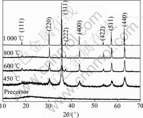
ͼ2 ��ͬ�����¶���������ά��Ʒ��XRD��
Fig.2 XRD patterns of fiber samples obtained at different calcination temperatures
��1 ��ͬ�¶����Ƶõ�Co0.5Ni0.5Fe2O4������ά��ƽ�������ߴ缰������ܲ���
Table 1 Average crystallite size (D) and magnetic parameters (Hc and Ms) of prepared Co0.5Ni0.5Fe2O4 nanofibers calcined at different temperatures
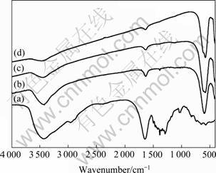
ͼ3 ��ά��Ʒ��FTIR��
Fig.3 FTIR spectra of fiber samples calcined at different temperatures: (a) Precursor fibers; (b) 450 ��; (c) 600 ��; (d) 800 ��
2.4 SEM��EDS����
ͼ4��ʾΪǰ������ά�����ڲ�ͬ�¶��±��� 2 h���ò����SEM���EDS�ס���ͼ4(a)~(e)���Կ����������¶ȶ���ά����ò�зdz����Ӱ�죬���ű����¶ȵ����ߣ���άֱ����С�������ھ��Ȼ����ֲ���Ϊ���У���ά������Խ���ֲڡ�ǰ������ά����������������������Ϊ�⻬������Ҳ��Ϊ���ȣ���άֱ���ֲ��Ͽ���Լ��500~2 000 nm֮�䡣��ͼ4(b)�ɼ�����450 �決�պ�����PVP�ͽ����εķֽ⡢��ά�ľ����Լ�Co0.5Ni0.5Fe2O4�����徧�����γɣ���άֱ�������С��Լ��200~400 nm֮�䣬��ά����Ҳ���Դֲڣ������ɼ�����֮�����γɵ�ϸС���������������������л����ȼ�ջӷ����ټ��ϴ�ʱ��ά�����ɵ�Co0.5Ni0.5Fe2O4�����徧����ΪϸС����ά���滹�����������������ά�ƺ����ֳ�һ�ֶ�ṹ�����ű����¶ȵĽ�һ������(��ͼ4(c)��(d))����άֱ����������С�����������徧���ij�����ά����Ŀ����Լ��٣����ܶ�����ߣ���ʱ��Ʒ����ά״�ṹ�Ա��ֽ�Ϊ��ã�������Ҳ�ȽϾ��ȡ����������¶����ߵ�1 000 ��ʱ����ͼ4(e)�ɼ�����ά��ò���������Եı仯�����ھ����Ĺ��ȳ���������ά�����ֳ�һ������״�ṹ����ʱ��ֱ����Ҫ�ֲ���150~300 nm��Χ�ڡ�
Ϊ�˽�һ����ʵ����ȷ�����Ʊ���Co-Ni������������ά�Ļ�ѧ��ɣ����о�����1 000 �決�����ɵ���ά��Ʒ��������ɫɢ����(��ͼ4(f))����������ɫɢ���У��۲쵽�˽���Ԫ��Fe��Co��Ni�ķ壬���ǵ�Ħ�������ֱ�Ϊ30.43%��7.84%��8.17%������Ŀ�����Co0.5Ni0.5Fe2O4������ά�Ļ�ѧ�����ȼ���ԭ����ȶ�����һ�¡�
2.5 TEM����
����TEM��һ���������Ʊ�����ά��Ʒ���۽ṹ��ͼ5��ʾΪ550��1 000 �決�պϳɵ�Co0.5Ni0.5Fe2O4������ά��TEM��ͼ5(a)���Կ�������550 �決���Ƶõ�Co0.5Ni0.5Fe2O4������ά�������С���������ۼ����ɣ���ƽ���ߴ�ԼΪ28.7 nm����������XRD���ݹ����ƽ�������ߴ�����൱����ͼ5(b)�ɼ����������¶���ߵ�1 000 ��ʱ�������ά�Ŀ����ߴ�������100 nm���ϣ�������ά�������ɵ���Co0.5Ni0.5 Fe2O4���������������ɣ��Ӷ��γ�һ�ֶ��ص�����״�����ṹ������ǰ��ķ���������Է��֣���ͬ�ı����¶ȵ������ɲ�ͬ�ߴ��������������ʹ��ά���ֳ���ͬ���۽ṹ����ò��
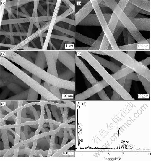
ͼ4 ��ά��Ʒ��SEM���EDS��
Fig.4 SEM images and EDS spectrum of as-prepared fiber samples: (a) Precursor fiber; (b) 450 ��; (c) 600 ��; (d) 800 ��; (e) 1 000 ��; (f) EDS spectrum in Fig.4(e)
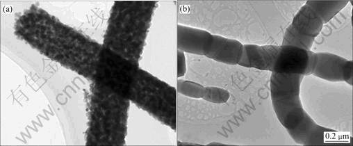
ͼ5 ��550��1 000 ��ϳɵ�Co0.5Ni0.5Fe2O4������ά�ĵ���TEM��
Fig.5 Typical TEM images of Co0.5Ni0.5Fe2O4 nanofibers synthesized at 550 �� (a) and 1 000 �� (b)
2.6 �����ܷ���
һ����������װ��ϵ�Ĵ���Ϊ����������ɿ�������ò(�ߴ����״)���仯������Ҳǿ�ҵ��ܵ�����������õ�Ӱ��[17]�����ִ��������Ҫ�����������䳤�̵�ż������ú�����������ԭ��֮��Ķ̳̽���������ã��ھ���ϵͳ�����������ϴ���һ����Ҫ�ĵ�λ��ͼ6��ʾΪ�����¶��±��պϳɵ�Co0.5Ni0.5Fe2O4������ά�����´��ͻ��ߡ���ͼ6�ɼ������ⳡԼΪ970 kA/mʱ����ʾ��ά��Ʒ�ĴŻ���δ�ﵽ���ͣ�ͨ������M��H����ʹ1/H�����㼴��M��Ľ���������Ʒ�ı��ʹŻ�ǿ��[31]�����¶������Ʊ���Co0.5Ni0.5Fe2O4������ά�ı��ʹŻ�ǿ��(Ms)�ͽ�����(Hc)����ǰ���1�С��ӱ�1���Կ�������ά�ı��ʹŻ�ǿ���決���¶ȵ��½�����С���� 1 000 ���80.2 A��m2/kg����С��450 ��ʱ��35.8 A��m2/kg��������ά��Co0.5Ni0.5 Fe2O4�����ijߴ��決���¶ȵı仯������ȫһ�¡�Co0.5Ni0.5Fe2O4������ά����Ӧ����������װ���ɣ���ˣ����ǵĴ�����������Co0.5Ni0.5Fe2O4�������������Լ�����֮�������á������о�����[9, 17, 32]�����ڳ�ϸ�����������Կ�������һ�������������еĺ˺�һ����������ԭ�ӴžصĿ�����ɣ��������������������ν�ķǹ����������ṹ����������״�ṹ������㣬�����ܹ��谭�����ڲ����˵���������ų��������С�����Co0.5Ni0.5Fe2O4�����ߴ���½����ȱ����������Դ�Ա���Ĺ������Եø�Ϊͻ�����Ӷ�������Ӧ��������άMs�ļ�С����Ʒ�Ľ������決���¶ȵı仯��Ϊ���Բ�ͬ�ڱ��ʹŻ�ǿ�ȣ��決���¶ȵ��½����ֳ���������С�����ƣ�Լ��550 ��ʱ�ﵽ���ֵ���������������ά�Ĵų�ṹת���й�[25]�����ű����¶ȵ��½�����ά��Co0.5Ni0.5 Fe2O4�����ijߴ���С����ų�ṹ�ɶ����ת�䣬��ת�����ϵĽ��������ﵽ���ֵ��ͨ���Խ��������ݽ�����Ϸ��֣�����ά��ʽ���ڵ�Co0.5Ni0.5Fe2O4���ṹ�Ĵŵ����ٽ�ߴ������30 nm���ҡ�
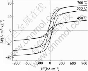
ͼ6 ���Ʊ�����ά��Ʒ�����´��ͻ���
Fig.6 Room-temperature hysteresis loops of as-prepared Co0.5Ni0.5Fe2O4 nanofiber samples
ͼ7��ʾΪ550 �決���Ƶõ�Co0.5Ni0.5 Fe2O4������ά������(RT)�͵���(77 K)�µĴ��ͻ��ߡ���ͼ7�п��Կ��������Ų����¶ȵ��½�����������ʣ��Ż�ǿ�Ⱦ��������ʹŻ�ǿ�ȼ�С��������ʱ��Ʒ�ı��ʹŻ�ǿ�ȡ�ʣ��Ż�ǿ�Ⱥͽ������ֱ�Ϊ64.5 A��m2/kg��17.9 A��m2/kg��72.1 kA/m������77 Kʱ���Ƿֱ�Ϊ44.6 A��m2/kg��33.4 A��m2/kg��618.2 kA/m�����Ƶ����������ù��������Ʊ���CoFe2O4��������ϵ��Ҳ���۲쵽[32]�������ڵ�������Ч����(��Ҫ�������������ܺ�������)������kBT�ı�ֵ���������´žص�ת�����������ˣ���77 Kʱ����Ʒ�ı��ʹŻ�ǿ�ȵ�������ֵ����������²��ִžصĶ���(������ת��)��ֱ�ӹ�ϵ��
����һ���ŵ������ṹϵͳ������������¶�(��������)�ı仯��Ϊ��������ʽ����������[33]��
 (2)
(2)
ʽ�У�Hc(0)�ǵ��������ھ������ʱ�Ľ�������
 (3)
(3)
ʽ�У�kB�Dz�������������T�Ǿ����¶ȣ�KE����Ч�Ÿ������Գ�����V�����������
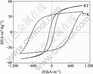
ͼ7 550 �決���Ƶõ�Co0.5Ni0.5Fe2O4������ά�����º͵����µĴ��ͻ���
Fig.7 Hysteresis loops at room temperature (RT) and low temperature (77 K) for prepared Co0.5Ni0.5 Fe2O4 nanofibers calcined at 550 ��
��ʽ(2)���Կ��������������¶ȵ��½��������ڵ����£��������Ŷ��Դ�ż����ת����Ӱ������������Ҫһ���������������ߵĴų����ı���ά����Щ�ʶ������е�ż���ӵĴŻ����Ӷ����º���ϱ��ֳ�һ������Ľ�����[34]��
Ϊ��̽���ڵ��뷶Χ����ά�Ľ�����(Hc)����ƽ�������ߴ�(D)�ľ���仯��ϵ����550 �����±��պϳɵ�Co0.5Ni0.5Fe2O4������ά��������ݽ�������Ϸ���(��ͼ8��ʾ)�������ڵ��뷶Χ�ڣ�Hc��D0.7�����ȣ���Hc��D0.7�����ڴ����ṹϵͳ����Ч������ϳ���Lex��������������ž��������Ե��½������ǵ�Lex���ܳ���ʵ����Ʒ�ijߴ磬�����ṹ���ϵ�ά�ȿ���Ҳ�Ǿ������������С��һ����Ҫ���������������������ģ�ͣ����Բ��ϵ���Ч�������Գ��� ����ά��m֮���������¹�ϵ[35]��
 (4)
(4)
ʽ�У�Kc�Dz��ϵĴž��������Գ�����A�ǽ�������ϵ����
ʽ(4)�������ڵ��뷶Χ�ڶ��ھ���һά(m=1)���ṹ�Ĵ��Բ��ϣ�������������侧���ߴ��2/3�η��仯����Hc��D2/3�����ڱ��о����Ʊ���Co0.5Ni0.5Fe2O4������ά��˵���ڵ����ٽ�ߴ����£���������ƽ�������ߴ�ı仯�Ϻõط���������Ԥ��Ľ������һ������˵���������������������������ʵ��ų�ṹ��Co0.5Ni0.5Fe2O4������ά�������µĴŻ��뷴�Ż���Ϊ����һ����Ҳ����ط�ӳ�����Բ��ϵ�ά��ȷʵ������������������Ӱ�졣
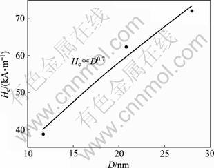
ͼ8 �����ٽ�ߴ�����Hc��D�ı仯
Fig.8 Variation of Hc with D as D below single-domain critical size for Co0.5Ni0.5Fe2O4 nanofibers
3 ����
1) ���þ����˿�����ɹ��Ʊ��⾧ʯ��Co0.5Ni0.5Fe2O4������ά�����ֱ����¶ȶ�������ά���۽ṹ����ò������Ҫ��Ӱ�졣���ű����¶ȵ����ߣ�ǰ������ά������̬����̬ת�䣬��450 �決��ʱ�����ྦྷ̬��Co0.5Ni0.5Fe2O4������ά�����γɡ�������ά���л�������εķֽ��Լ������徧���γɺͳ������±��պ���άֱ����С�������ø�Ϊ�ֲں����ܣ�������ò��������״�ṹת�䡣��1 000 �決��2 h���Ƶõ�Co0.5Ni0.5Fe2O4������ά������״�ṹ����ֱ��Լ��150~300 nm֮�䡣
2) �決���¶ȵ����ߣ����Ʊ���Co0.5Ni0.5Fe2O4������ά�ı��ʹŻ�ǿ��������������������������С�����ƣ���550 �渽���ﵽ���ֵ��Co0.5Ni0.5Fe2O4������ά�ĵ����ٽ�ߴ�ԼΪ30 nm���ڵ��뷶Χ�ڣ�������(Hc)��ƽ�������ߴ�(D)��0.7�η������ȣ��Ϻõط��������������ģ����Ԥ��Ľ����
3) �ڵ����£��������Ŷ�Ӱ��ļ����Ͳ��ִžصĶ��ᣬ��ά�Ľ�������ʣ��Ż�ǿ����������¾��д����ߣ����ʹŻ�ǿ�ȳ���һ���̶ȵ��½���
REFERENCES
[1] �Ų���, �� ��, �� ÷, ������, �� ��, ���. �����ϲ���Co0.5Ni0.5Fe2O4-SiO2 �����ṹ�ʹ���[J]. ������ѧ��, 2008, 36(3): 292-295.
ZHANG Bo-jun, HUA Jie, LIU Mei, XU Shi-chong, FENG Ming, LI Hai-bo. Microstructures and magnetic properties of Co0.5Ni0.5Fe2O4-SiO2 nanocomposites[J]. Journal of the Chinese Ceramic Society, 2008, 36(3): 292-295.
[2] Pankhurst Q A, Thanh N K T, Jones S K, Dobson J. Progress in application of magnetic nano-particles in biomedicine[J]. J Phys D: Appl Phys, 2009, 42: 22401.
[3] Mathe V L, Kamble R B. Anomalies in electrical and dielectric properties of nanocrystalline Ni-Co spinel ferrite[J]. Mater Res Bull, 2008, 43: 2160-2165.
[4] �� ��, �� ̩, ��Ӣ, �� ��. ���� Co1�CxNixFe2O4��������Ʊ���Ni2+��������ܵ�Ӱ��[J]. ������ѧ��, 2007, 35(2): 160-163.
LIU Yin, QIU Tai, SHEN Chun-ying, YANG Jian. Preparation of nanocrystalline Co1�CxNixFe2 O4 ferrite and effect of Ni2+ on its magnetic properties[J]. Journal of the Chinese Ceramic Society, 2007, 35(2): 160-163.
[5] Song Q, Zhang Z J. Shape control and associated magnetic properties of spinel cobalt ferrite nanocrystals[J]. J Am Chem Soc, 2004, 126: 6164-6168.
[6] Kim D H, Nikles D E, Johnson D T, Brazel C S. Heat generation of aqueously dispersed CoFe2O4 nanoparticles as heating agents for magnetically activated drug delivery and hyperthermia[J]. J Magn Magn Mater, 2008, 320: 2390-2396.
[7] Niu Z P, Wang Y, Li F S. Magnetic properties of nanocrystalline Co-Ni ferrite[J]. J Mater Sci, 2006, 41: 5726-5730.
[8] Gul I H, Amin F, Abbasi A Z, Anis-ur-Rehman M, Maqsood A. Physical and magnetic characterization of co-precipitated nanosize Co-Ni ferrites[J]. Scripta Mater, 2007, 56: 497-500.
[9] Maaz K, Khalid W, Mumtaz A, Hasanain S K, Liu J, Duan J L. Magnetic characterization of Co1-xNixFe2O4 (0��x��1) nanoparticles prepared by co-precipitation route[J]. Physica E, 2009, 41: 593-599.
[10] Kasapoglu N, Birs?z B, Baykal A, K?seoglu Y, Toprak M S. Synthesis and magnetic properties of octahedral ferrite NixCo1-xFe2O4 nanocrystals[J]. Cent Eur J Chem, 2007, 5: 570-580.
[11] Jiang J. A facile method to the Ni0.8Co0.2Fe2O4 nanocrystalline via a refluxing route in ethylene glycol[J]. Mater Lett, 2007, 61: 3239-3242.
[12] Shobana M K, Rajendran V, Jeyasubramanian K, Kumar N S. Preparation and characterisation of NiCo ferrite nanoparticles[J]. Mater Lett, 2007, 61: 2616-2619.
[13] Singhal S, Singh J, Barthwal S K, Chandra K. Preparation and characterization of nanosize nickel-substituted cobalt ferrites (Co1-xNixFe2O4)[J]. J Solid State Chem, 2005, 178: 3183-3189.
[14] Zhang F, Kitamoto Y, Abe M, Naoe M. Effect of Ni addition into Co ferrite thin films for perpendicular recording media[J]. J Appl Phys, 2000, 87: 6881-6883.
[15] Bayrakdar H, Esmer K. Dielectric characterization of NixCo1-xFe2O4 nanocrystals thin film over a broad frequency range (1 MHz-3 GHz)[J]. J Appl Phys, 2010, 107: 044102.
[16] Allwood D A, Xiong G, Cooke M D, Faulkner C C, Atkinson D, Vernier N, Cowburn R P. Submicrometer ferromagnetic not gate and shift register[J]. Science, 2002, 296: 2003-2006.
[17] Wang Z L, Liu X J, Lv M F, Chai P, Liu Y, Zhou X F, Meng J. Preparation of one-dimensional CoFe2O4 nano- structures and their magnetic properties[J]. J Phys Chem C, 2008, 112: 15171-15175.
[18] Li D, Herricks T, Xia Y N. Magnetic nanofibers of nickel ferrite prepared by electrospinning[J]. Appl Phys Lett, 2003, 83: 4586-4588.
[19] ��ΰ��, �¿���, ���, ����Ƽ. ����ֲ�Fe�����߸��ϲ��ϵ���������[J]. �й���ɫ����ѧ��, 2005, 15(2): 288-294.
PENG Wei-cai, CHEN Kang-hua, LI Jing-lei, HUANG Lan-ping. Microwave absorbing properties of iron nanowire composites distributed randomly[J]. The Chinese Journal of Nonferrous Metals, 2005, 15(2): 288-294.
[20] Li D, McCann J T, Xia Y N. Electrospinning: A simple and versatile technique for producing ceramic nanofibers and nanotubes[J]. J Am Ceram Soc, 2006, 89: 1861-1869.
[21] Ramaseshan R, Sundarrajan S, Jose R, Ramakrishna S. Nanostructured ceramics by electrospinning[J]. J Appl Phys, 2007, 102: 111101.
[22] Tan S T, Huang X W, Wu B L. Some fascinating phenomena in electrospinning processes and applications of electrospun nanofibers[J]. Polym Int, 2007, 56: 1330-1339
[23] Ju Y W, Park J H, Jung H R, Cho S J, Lee W J. Electrospun MnFe2O4 nanofibers: Preparation and morphology [J]. Compos Sci Technol, 2008, 68: 1704-1709.
[24] Maensiri S, Sangmanee M, Wieng-moon A. Magnesium ferrite (MgFe2O4) nanostructures fabricated by electrospinning[J]. Nanoscale Res Lett, 2009, 4: 221-228.
[25] Xiang J, Shen X Q, Song F Z, Liu M Q. Fabrication and magnetic properties of Ni0.5Zn0.5Fe2O4 nanofibers by electrospinning[J]. Chin Phys B, 2009, 18: 4960-4965.
[26] Xiang J, Shen X Q, Song F Z, Liu M Q. One-dimensional NiCuZn ferrite nanostructures: Fabrication, structure, and magnetic properties[J]. J Solid State Chem, 2010, 183(6): 1239-1244.
[27] �� ��, �θ�չ, ����ǭ, ������. һάNi0.5Zn0.5Fe2O4/SiO2�������ṹ���Ʊ����������[J]. ����ѧ��, 2010, 59(7): 4794-4801.
XIANG Jun, SONG Fu-zhan, SHEN Xiang-qian, CHU Yan-qiu. Preparation of one-dimensional Ni0.5Zn0.5Fe2O4/SiO2 composite nanostructures and their magnetic properties[J]. Acta Physica Sinica, 2010, 59(7): 4794-4801.
[28] ������, �� ��, ����͢, ����ϼ. ZnO@SiO2ͬ�������µľ����˿�����Ʊ������[J]. ����ѧѧ��, 2010, 26(1): 29-34.
WANG Jin-xian, ZHANG He, DONG Xiang- ting, LIU Gui-xia. ZnO@SiO2 coaxiel nanocable: preparation via electrospinning and characterization[J]. Chinese Journal of Inorganic Chemistry, 2010, 26(1): 29-34.
[29] Priyadharsini P, Pradeep A, Sambasiva Rao P, Chandrasekaran G. Structural, spectroscopic and magnetic study of nanocrystalline Ni-Zn ferrites[J]. Mater Chem Phys, 2009, 116: 207-213.
[30] Yan H W, Zhang K, Blanford C F, Francis L F, Stein A. In vitro hydroxycarbonate apatite mineralization of CaO-SiO2 sol-gel glasses with a three-dimensionally ordered macroporous structure[J]. Chem Mater, 2001, 13(4): 1374-1382.
[31] Dolia S N, Prasad A S, Dhawan M S, Sharma M P, Chander S. Magnetic behaviour of nanocrystalline Ni0.5Cu0.5Fe2O4 spinel ferrite[J]. Hyperfine Interact, 2008, 184: 75-81.
[32] Mumtaz A, Maaz K, Janjua B, Hasanain S K, Bertino M F. Exchange bias and vertical shift in CoFe2O4 nanoparticles[J]. J Magn Magn Mater, 2007, 313: 266-272.
[33] ���ž�, ������, ����ѫ, �Ž��. ��п�����屡Ĥ�����ṹ�͵��´�����[J]. ���ܲ���, 2005, 36(12): 1855-1858.
WANG Jiu-jing, YU Li-ming, CAO Shi-xun, ZHANG Jin-cang. Microstructure and low temperature magnetic properties of NiZn-ferrite film[J]. Journal of Functional Materials, 2005, 36(12): 1855-1858.
[34] Wu H, Zhang R, Liu X X, Lin D D, Pan W. Electrospinning of Fe, Co, and Ni nanofibers: Synthesis, assembly, and magnetic properties[J]. Chem Mater, 2007, 19: 3506-3511.
[35] Sellmyer, D J, Liu Y, Shindo, D. Handbook of advanced magnetic materials (Vol. 1) Advanced magnetic materials: Nanostructural effects[M]. Beijing: Tsinghua University Press, 2005: 354.
(�༭ ����)
������Ŀ��������Ȼ��ѧ����������Ŀ(50674048)������ʡ��ͨ��У�о������д��¼ƻ�������Ŀ(CX09B-192Z)������ʡ��������������Ŀ�����տƼ���ѧ����Ǹɽ�ʦ֧�ּƻ�������Ŀ
�ո����ڣ�2010-08-31�������ڣ�2011-01-24
ͨ�����ߣ��� ���������ڣ���ʿ���绰��15952808679��E-mail: junx93@sina.com

